3933
Functional diffusion maps to assess treatment response in head and neck tumors using SPLICE.1Radiotherapy, Universtiy Medical Center Utrecht, Utrecht, Netherlands, 2Cancer Center, Universtiy Medical Center Utrecht, Utrecht, Netherlands
Synopsis
ADC changes during chmoradiation treatment might be of prognostic value in patients with head and neck squamous cell carcinoma (HNSCC) and allow for treatment modification. To overcome decrease in tumor visibility and increase in delineation variation by observers, functional diffusion maps provide an objective measure to follow response on DW-MRI during treatment provided a good geometric accuracy as is offered by the SPLICE technique.
Introduction
The apparent diffusion coefficient (ADC) determined by diffusion weighted magnetic resonance imaging (DW-MRI) is a surrogate measurement of cellularity and stromal component of a tumor. (Chemo)radiation treatment can result in ADC increase and decrease depending on the timing and the individual tissue response. ADC changes during treatment might be of prognostic value in patients with head and neck squamous cell carcinoma (HNSCC) and allow for treatment modification. However, tumor visibility on MRI decreases during treatment hampering tumor delineation and increasing observer variation. To avoid this, functional diffusion maps can be used to objectively analyze ADC changes in time on a per-voxel basis (1). This requires images with good geometric accuracy to enable image registration of the time series. Split-echo acquisition of FSE signal (SPLICE) sequences have superior geometric performance to EPI sequences and can therefore provide these distortion free DW-MRI images (2,3).Purpose
To follow treatment response on DWI of head and neck tumors using functional diffusion maps.Methods
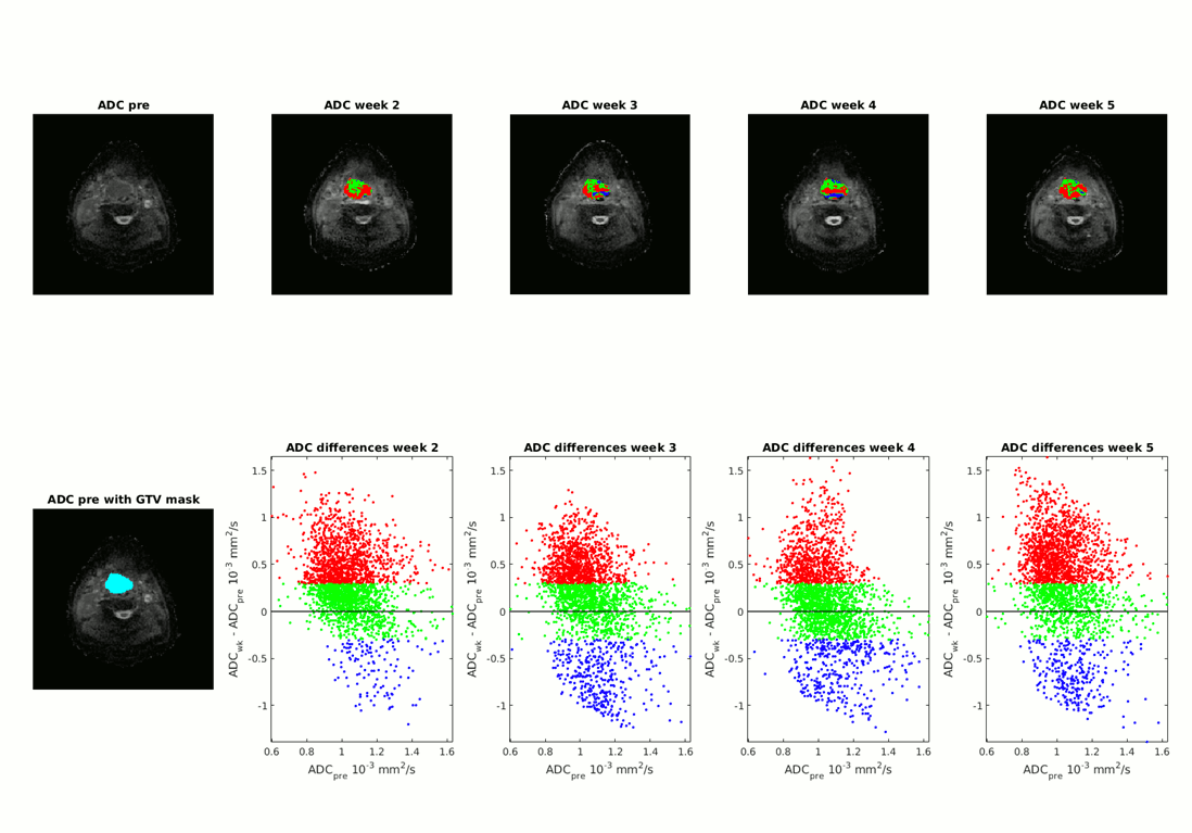
Twelve patients treated with radiotherapy, with or without concurrent chemotherapy, underwent MRI prior to and during week 2, 3, 4 and 5 of the radiotherapy treatment. Imaging was obtained with the patient positioned in the radiotherapy mask. The MRI protocol contained a SPLICE DW-MRI sequence with b-values of 0, 200 and 800 s/mm2. Acquisition was performed using the following sequence parameters: TR 16366 ms, TE 52 ms, SENSE 2, echo train length 64, pixel bandwidth 900 Hz, FOV (RL x AP ) 230 x 280 mm2 (FH) 120 mm, acquired voxel size 1.8 x 1.8 mm2, slice thickness 4 mm. Image registration was performed on the b=0 image of teh SPLICE seqeunce using mutual information. The tumor was solely delineated on the pretreatment diffusion weighted MR images and ADC map resulting in a tumor mask. ADC maps were all registered rigidly to the MRI pretreatment. Functional diffusion maps were created for all patients using a threshold of 0.3 10-3 mm2/s for ADC changes (figure 1).Results
During (chemo)radiotherapy more tumor voxels showed increase (mean 33%) in ADC than a decrease (9%) (figure 2). ADC increase was already apparent in the second week of treatment except in one patient. ADC decrease mostly points to volume decrease, which was most apparent from week 4 during treatment on.Discussion
After (chemo)radiation treatment, contouring of a tumor is difficult due to radiation induced changes in the tumor and surrounding tissue including edema, apoptosis, necrosis and fibrosis induction. Diffusion weighted image acquisition in radiotherapy mask with the SPLICE method allows for accurate registration of the time series of diffusion weighted images due to very limited image distortions unlike common EPI based diffusion weighted imaging. This enabled response assessment on diffusion weighted imaging using the functional diffusion maps in this region with large susceptibility differences. As the follow up period of these patients is still limited, the clinical evaluation of the functional diffusion maps will be performed in a later stage.Conclusion
During (chemo)radiotherapy for HNSCC, tumor response using DW-MRI mostly showed increase of apparent diffusion coefficient in around one third of voxels as soon as the second week into treatment. This can be interpreted as reduction of cell density by necrosis, replacement of tumor by normal tissue and edema in and around remaining tumor tissue. Decrease in ADC is most evident from week 4 into treatment on. This suggests a reduction of tumor voxels replaced by air containing voxels. Functional diffusion maps provide an objective measure to follow response on DW-MRI during treatment provided a good geometric accuracy as is offered by the SPLICE technique.Acknowledgements
No acknowledgement found.References
[1] Hamstra D, Chenevert TL, Moffat B, Johnson TD, Meyer CR, Mukherji SK, et al. Evaluation of the functional diffusion map as an early biomarker of time-to-progression and overall survival in high-grade glioma. Proc Natl Acad Sci U S A. 2005;102(46):16759–64.
[2] Schick F. SPLICE: sub-second diffusion-sensitive MR imaging using a modified fast spin-echo acquisition mode. Magn Reson Med. 1997 Oct;38(4):638-44.
[3] Schakel T, Hoogduin JM, Terhaard CHJ, Philippens MEP. Technical Note: Diffusion-weighted MRI with minimal distortion in head-and-neck radiotherapy using a turbo spin echo acquisition method. Med Phys. 2017 Aug;44(8):4188-4193.
Figures