3914
Radiomics: a novel MRI-based method of predicting recurrence in chordoma1Key Laboratory of Molecular Imaging, Chinese Academy of Science, Beijing, China, 2School of Life Sciences and Technology, Xidian University, Xi'an, China, 3Department of Electronics and Information, Xi'an Polytechnic University, Xi'an, China, 4Department of Neurosurgery, Beijing Tiantan Hospital, Capital Medical University, Beijing, China
Synopsis
In order to find the relationship between MRI image and the postoperative recurrence of chordoma, we used a novel radiomics method for quantitative analysis of MRI image. Finally successfully predicted the probability of postoperative recurrence of chordoma.
Authors
Wei Wei, Ke Wang and Kaibing Tian contributed equally to this work.Corresponding author: Jie Tian, jie.tian@ia.ac.cn. Zhen Wu, wuzhen1966@aliyun.com.Introduction
Chordoma is a rare primary malignant tumor1, which originated from residual embryos, and the annual incidence rate was only 0.5 to 0.8 / million2. The local recurrence rate was as high as 43% to 85%, and most recurrences occurred in 3 years after operation3.For evaluating the related factors of postoperative recurrence, we develop and internally validate a radiomics model, along with routinely available MRI, to identify patients who would recurrence, and identify MRI-based radiomics features as prognostic factors in patients with chordoma4.Patients And Methods
Our cohort included 155 patients with chordoma from Beijing Tiantan Hospital, Capital Medical University. All patients had complete follow-up data and T1-weighted image (T1WI), T2-weighted image (T2WI) and contrast-enhanced T1-weighted (CET1-w) MRI sequence data. All manual segmentations of the region of interest (ROI, tumor) were performed by a radiologist who had 10 years of experience, and each segmentation was validated by a senior radiologist, who had 20 years of experience. 358 radiomics features (First order statistics features, Shape and size based features, Textural features, Wavelet features) were extracted from T1WI, T2WI and CET1-w MRI, respectively. Calculated each feature and the predictive value of the corresponding Area Under roc Curve (AUC) value for single feature analysis, and then selected the feature with AUC value greater than 0.5 as candidate feature. Three sequences were obtained a set of candidate features respectively, and used Elastic Net analysis to construct prediction model5. The value of alpha was set to 0.1, the minimum value of lambda was set to 0.00001, and the number of lambda was set to 100. The model used 10-fold cross validation, and used AUC value as the cross validation judgment standard, predict and analysis based on the model corresponding to lambda_1se value. Logistic regression classifier predicted the recurrence probability.Results
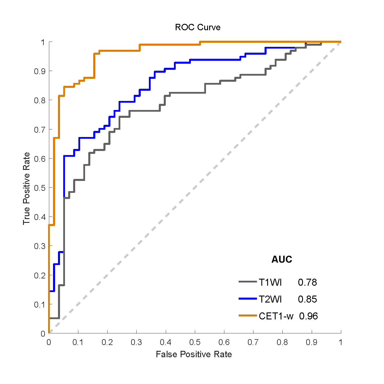
T1WI model integrates 21 features. The AUC value corresponding to the Receiver Operating Characteristic (ROC) curve was 0.78, the accuracy (ACC) was 0.71. T2WI model integrates 21 features. The AUC value corresponding to the ROC curve was 0.85, the ACC was 0.79. CET1-w model integrated 79 features. The AUC value corresponding to the ROC curve was 0.96, the ACC was 0.92 (Figure 1). According to Elastic Net- Logistic regression analysis, T1WI, T2WI and CET1-w models were significantly associated with recurrence among the 155 patients.Discussion
To our knowledge, this was the first study based on the quantitative analysis of MRI image radiomics method to be applied in the prediction of the recurrence in chordoma. Radiomics features were extracted from three different MRI sequences (T1WI, T2WI and CET1-w sequence). Through Elastic Net- Logistic regression modeling analysis, three models successfully divided patients into high-risk and low-risk groups, and the CET1-w sequence prediction ability was superior to T1WI and T2WI sequences. CET1-w sequence and chordoma recurrence was most closely linked. There are still limitations to our study. It is a small sample of retrospective study, and all cases come from a single institute, thus reducing the generalization ability of the model.Conclusion
Radiomics model based on MRI and quantitative image features provided prognostic ability in chordoma. The models are closely associated with recurrence, especially the CET1-w model. These results provide an illustrative example of precision medicine and may affect treatment strategies.Acknowledgements
No acknowledgement found.References
1. McMaster ML, Goldstein AM, Bromley CM, et al. Chordoma: incidence and survival patterns in the United States, 1973–1995. Cancer Causes Control. 2001; 12:1–11.
2. Eriksson B, Gunterberg B, Kindblom LG. Chordoma: aclinicopathologic and prognostic study of a Swedish nationalseries. Acta Orthop Scand. 1981; 52:49–58.
3. Yang YK, Niu XH, Li Y, et al. Recurrence and survival factors analysis of 171 cases of sacral chordoma in a single institute. European Spine Journal. 2017; 26(7):1910-1916.
4. Aerts HJ, Velazquez ER, Leijenaar RT, et al. Decoding tumour phenotype by noninvasive imaging using a quantitative radiomics approach. Nat Commun. 2014; 5:4006.
5. Zou H, Hastie T. Regularization and variable selection via the elastic net. Journal of the Royal Statistical Society Series B-Statistical Methodology.2005; 67:301-320.
Figures