3890
Changes of brain metabolites in response to acupuncture therapy in migraine are correlated with clinical outcomes: Result from a magnetic resonance spectroscopy imaging Study1Beijing hospital, Beijing, China, 2ImageTech laboratory, Simon Fraser University, Surrey, BC, Canada
Synopsis
Migraine is a common neurological disease. Acupuncture has been proven to be effective but the mechanisms remain unclear. A proton magnetic resonance spectroscopy imaging (MRSI) study was used to investigate biochemical changes in brain regions key for the transmission of pain in response to acupuncture treatment. Results showed acupuncture treatment was associated with a significantly increased NAA/Cr in bilateral thalamus in migraine patients. A strong significant correlation between NAA/Cr and headache intensity was found in thalami. Our data provided the first evidence suggesting a brain biochemical change in response to acupuncture therapy in migraine, in correlation with clinical outcomes.
Introduction
Migraine is a common neurological disease with a high prevalence and unsatisfactory treatment options. Acupuncture has been proven to be effective but the specific pathophysiological mechanisms remain unclear, limiting the development of the treatment. Here we conducted a proton magnetic resonance spectroscopy imaging (MRSI) study, aiming to investigate biochemical changes in brain regions key for the transmission of pain in response to acupuncture treatment. We also linked the brain metabolite changes with clinical headache assessment for patients.Material and Methods
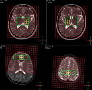
Participants included patients diagnosed as having migraine with no aura patients (MnA) or cervical headache (CH) and normal controls (NC); n=15 in each group. All participants received acupuncture treatment (verum or sham) and had MRSI scans before and after the treatment. 2D multiple voxel PRESS sequence was applied to acquire spectrum from three targeted VOIs (Fig. 1), positioned within the bilateral thalamus, anterior cingulate cortex and posterior part of paracentral gyrus areas. Brain metabolites such as N-acetylaspartate (NAA), creatine (Cr) and choline compounds (Cho) were examined together with headache intensity, headache frequency and duration of headache attack evaluations.Results
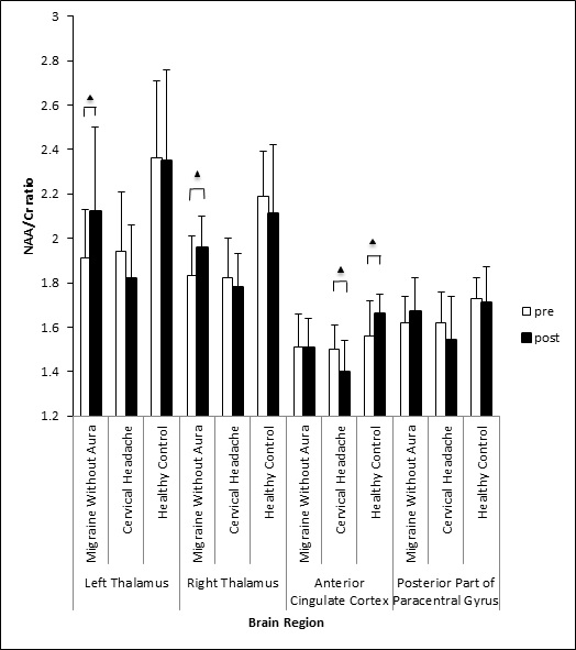
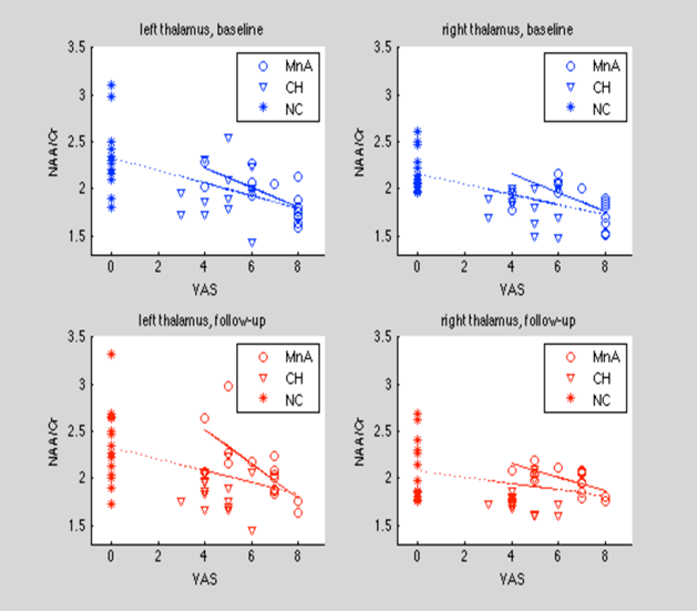
Acupuncture treatment was associated with a significantly increased NAA/Cr in bilateral thalamus in MnA patients, which was not observed in the CH or NC groups (Fig. 2). A significant correlation between NAA/Cr and headache intensity was found in both the left and the right thalamus for all participants, while the correlations were stronger in MwoA patients (Fig. 3).Discussion
The study is the first of the kind in investigating the effect of acupuncture therapy on brain metabolites in migraine using magnetic resonance spectroscopy imaging. The study provides the initial result evidencing a positive response of brain metabolites to acupuncture therapy. Our data showed that indeed, an increase of NAA/Cr (while Cr remained relatively stable) in bilateral thalamus was achieved through acupuncture therapy of just 5 sessions, associated with a decrease in headache severity. NAA is generally recognized as a marker of normal neuronal activity, synthesized/located in neuronal mitochondria; therefore NAA decrease indicates mitochondrial dysfunction [1]. Thus, such an increase in NAA likely reflects a recovery of neuronal activity in the thalamus through mediating nociceptive modulation [2].The ACC, on the other hand, is involved in the emotional response to pain (affective aspects of pain perception), as well as in a behavior and reinforcement response to pain [3]. This provides an explanation why in the CH group, the acupuncture procedure did not benefit thalamic modulation but can also be harmful (i.e., a decrease of NAA/Cr in the ACC in the CH group), suggesting the importance of applying the treatment on the effective acupoints based on the TCM theory, for the targeted disease or condition [4]. By the same token, in the NC group, applying the acupuncture procedure at non-acupoint spots did not generate an effect as effective as in the MnA group. Similarly, a change of the metabolites was not found in the PPG in any group. Even though PPG is involved in encoding of the location, intensity and duration, it is not involved in the modulation of headache, as neurons in this region receive nociceptive signals from body and limbs, not head and face [5]. The correlations between the metabolites of interest with clinical measures demonstrated the clinical mindfulness of MRSI findings in MwoA group. Maintaining a sufficient level of the metabolites is necessary to ease headache symptoms. Improvement of migraine outcome was related with the increase of the thalamic NAA level in the patient group, which received the true treatment.Conclusion
Our data provided the first evidence suggesting a brain biochemical change in response to acupuncture therapy in migraine, in correlation with clinical outcomes. It also demonstrated the utilization of MRSI in better understanding the mechanisms of acupuncture treatment of migraine.Acknowledgements
This study was supported by grants from the National Natural Science Foundation of China (30901928), Capital’s Funds for Health Improvement and Research (2014-4-4052), and the Surrey Hospitals and Outpatient Centre Foundation (G2017-001).References
1. Clark JB. N-acetyl-aspartate: a marker for neuronal loss or mitochondrial dysfunction. Dev Neurosci 1998, 20(4-5): 271–6. 2. Tang JS, Qu CL, Huo FQ. The thalamic nucleus submedius and ventrolateral orbital cortex are involved in nociceptive modulation: A novel pain modulation pathway. Prog Neurobiol, 2009, 89(4):383-89. 3. Rushworth MF, Behrens TE, Rudebeck PH, Walton ME: Contrasting roles for cingulate and orbitofrontal cortex in decisions and social behaviour. Trends Cogn Sci, 2007, 11(4):168-76. 4. State Administration of Traditional Chinese Medicine. The criterion of diagnosis and therapeutic effects of disease and syndrome in traditional Chinese medicine. Nanjing University Press, 1994, p22-23. 5. Lim SH, Dinner DS, Pillay PK, et al. Functional anatomy of the human supplementary sensorimotor area: results of extraoperative electrical stimulation. Electroencephaloqr Clin Neurophysiol, 1994, 91(3):179-93.Figures