3887
Quintuple tuned coil configuration for multi-nuclear metabolic MRI at 7 tesla1Radiology, University Medical Center Utrecht, Utrecht, Netherlands, 2MR Coils, Zaltbommel, Netherlands
Synopsis
Metabolic and anatomic imaging by combining X-nuclei imaging with 1H imaging has shown great potential in clinical research. Traditional multi-nuclear coil setups are however limited to 2 or 3 frequencies, and often birdcage for 1H. In this study we propose a coil array setup tuned for acquiring 5 different nuclei in a single scan session practically without compromising efficiency on any nuclei.
Introduction
Metabolic imaging with MRI can provide a wide variety of information. Particular when non-proton nuclei are observed during the MR exam. For example: tumor response to chemotherapy, tracking a variety of metabolic pathways, analyzing the state of cartilage and the conversion of drugs in the body can be observed with 19F, 31P, 23Na and 13C MRI. However, generally only one or two nuclei besides 1H can be observed during a single examination1, often with limited performance on 1H.
Here we present a quintuple tuned coil setup for 7T that can be realized practically without compromising performance on any nucleus due to tissue load dominance. The proposed setup consists of a triple tuned birdcage transmit coil, combined with a triple tuned local receiver loop array, all tuned for the 13C, 23Na and 31P nuclei. These loops can be combined with broadband antennas for transceiving 19F and 1H as an array. This setup enables observing 5 different nuclei – 1H, 19F, 31P, 23Na and 13C – at 7 tesla within a single examination.
Methods
A birdcage transmit/receive coil designed for the extremities was tuned on the frequencies of 13C, 23Na and 31P at 7 tesla (75, 79 and 121 MHz). Positioned inside the birdcage were 4 triple tuned receive-only loops for the same frequencies (MR Coils BV, Zaltbommel, NL). On top of the receiver-array, a broadband transmit/receive antenna for 19F and 1H (280 and 298 MHz) was positioned2. Note that the tuning from 75 to 79 MHz is realized by an active PIN diode switch.
Bench experiments:
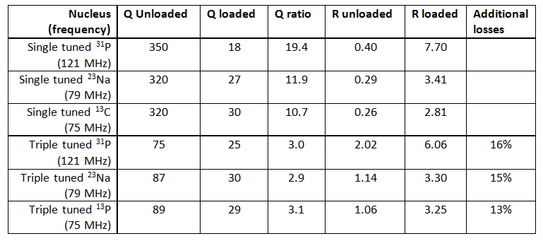
To determine the
losses when moving from single to triple tuned receiver-coils, Q ratios were
determined with a network analyzer. For this, three single tuned coils were
built for each nucleus as well as the triple tuned coil. Unloaded and loaded Q values
of different subjects were measured. Additional losses in SNR by going from
single to triple tuned coils were calculated based on the loaded to unloaded
conditions.
In-vivo experiments: All experiments were performed on the lower arm of a healthy volunteer in a whole body 7 tesla MR system (Philips, Best, NL).
Due to current interfacing constraints of the scanner platform, the receiver array could not be used for 23Na and 13C in-vivo experiments. For these nuclei, reception of the signals was done with the birdcage coil instead. Further, due to the low natural abundance of 19F in the body, no 19F experiments were performed in this study.
For the in-vivo experiments, without any B0 shimming, the following fast scan parameters were used:
- 13C: SV MRS, 4096 data points, 512 averages, BW: 16000Hz, 205s
- 23Na: SV
MRS, 512 data points, 1024 averages, BW: 16000Hz, 41s
- 31P: SV
MRS, 512 data points, 30 averages, BW: 6000Hz, 150s
- 1H: 3D FFE, FA: 45°, TE/TR: 3/25ms, 2x2x3mm3, FOV: 120x500x174mm3, 7s
- Total scan time: 7minutes
Results
Bench experiments: Q-ratios of Rx loops decreased with a factor of 3.5 to 6.5 when going from single tuned to triple tuned coils (table 1).
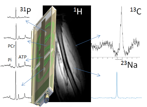
In-vivo experiments: Figure 1 shows spectra of 3 different nuclei and a proton image, acquired with a single setup. The phosphorous experiments clearly show phosphocreatine, ATP and inorganic phosphate at high SNR. Sodium and carbon signals are shown as well
Discussion and conclusion
In this study we presented a quintuple tuned coil setup, enabling MR experiments of 5 different nuclei in a single examination. The setup consists of a triple tuned birdcage coil for transmitting and receiving of signals on 3 frequencies, combined with antennas for 2 frequencies. A local receiver array was constructed for the lower 3 frequencies.
Tuning the receiver coils for multiple nuclei does have a penalty in terms of SNR. However, these types of coils are placed in an receiver array. This can result in an increase in SNR and enables the use of parallel imaging techniques3,4. Moreover, the transmit coil provides a uniform field avoiding the need for adiabatic RF pulses.
As our scanner platform currently is limited to one receiver for frequencies lower than 120MHz, only 31P experiments could be performed with the receiver array. As the Q-factor ratio of all three nuclei are similar, we expect similar performance from the 13C and 23Na frequencies.
No 19F experiments were demonstrated in this work. However, previous research shows that antennas similar as used in this study are capable of detecting 19F containing drugs in-vivo2.
While here we demonstrated only a single antenna with 4 loops in a small birdcage, the concept would be scalable to a multi-transmit setup with many more receivers in a whole body birdcage.
To conclude, quintuple tuned coil setups are a feasible option to enable multi-nuclear metabolic MRI at 7 tesla.
Acknowledgements
No acknowledgement found.References
1. Lee, R. F., Giaquinto, R., Constantinides, C., Souza, S., Weiss, R. G. and Bottomley, P. A. (2000), A broadband phased-array system for direct phosphorus and sodium metabolic MRI on a clinical scanner. Magn. Reson. Med., 43: 269–277. doi:10.1002/(SICI)1522-2594(200002)43:2<269::AID-MRM14>3.0.CO;2-J
2. van Gorp, J. S., Seevinck, P. R., Andreychenko, A., Raaijmakers, A. J. E., Luijten, P. R., Viergever, M. A., Koopman, M., Boer, V. O., and Klomp, D. W. J. (2015) 19 F MRSI of capecitabine in the liver at 7 T using broadband transmit–receive antennas and dual-band RF pulses. NMR Biomed., 28: 1433–1442. doi: 10.1002/nbm.3390.
3. Roemer, P. B., Edelstein, W. A., Hayes, C. E., Souza, S. P. and Mueller, O. M. (1990), The NMR phased array. Magn Reson Med, 16: 192–225. doi:10.1002/mrm.1910160203
4. Pruessmann, K. P., Weiger, M., Scheidegger, M. B. and Boesiger, P. (1999), SENSE: Sensitivity encoding for fast MRI. Magn. Reson. Med., 42: 952–962. doi:10.1002/(SICI)1522-2594(199911)42:5<952::AID-MRM16>3.0.CO;2-S
Figures