3881
Coil Combination Methods for 16-channel Hyperpolarized 13C Spectroscopic Imaging Studies of Liver Metastases Patients1Department of Radiology and Biomedical Imaging, UCSF, San Francisco, CA, United States, 2UC Berkeley - UCSF Graduate Program in Bioengineering, UCSF, San Francisco, CA, United States, 3Department of Medicine, UCSF, San Francisco, CA, United States
Synopsis
Effective coil combination methods for human hyperpolarized 13C spectroscopy data remain relatively unexplored. This study implemented several coil combination methods, including sum-of-squares (SOS), singular value decomposition (SVD), and pyruvate map based sensitivity calibration (PyrMap). These methods were evaluated by both simulation and in human cancer studies. Overall, both the SVD and PyrMap methods demonstrated better accuracy and robustness than SOS, and the PyrMap best preserved the phase information.
Purpose
With the recent advances on hyperpolarized 13C clinical studies, the safety, efficacy, and unique metabolic information provided by this technique have been demonstrated1. The use of multi-channel coils is necessitated due to the need of whole-body coverage and the limited lifetime of hyperpolarized signals. The goals of this study were to investigate and evaluate coil combination methods, and to develop a robust framework for multi-channel clinical hyperpolarized 13C spectroscopy data combination.Methods
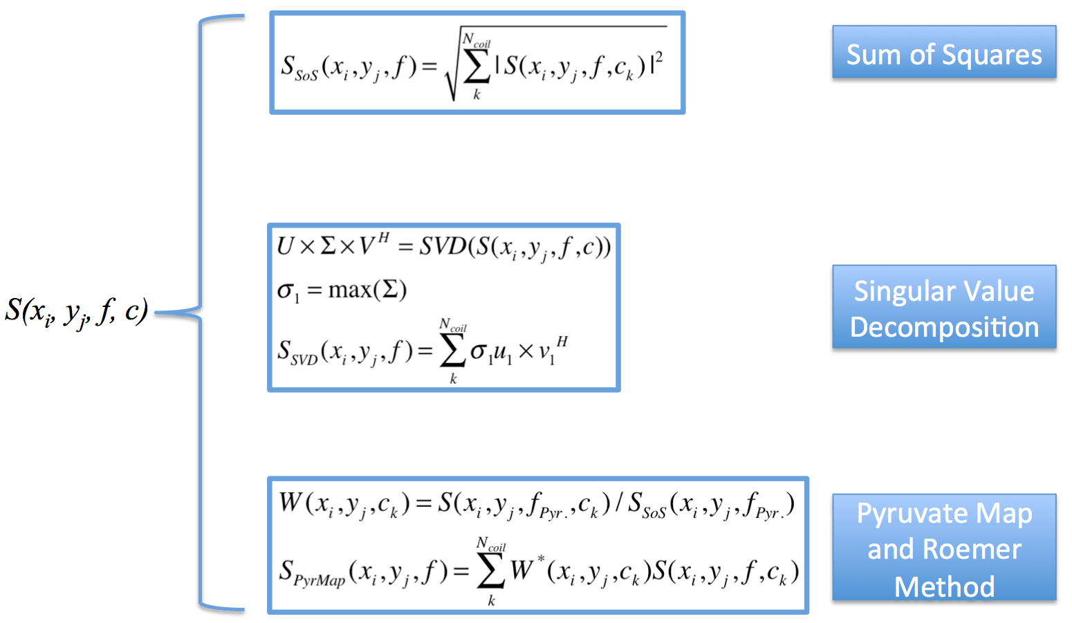
Three coil combination methods were evaluated in this study, namely the sum-of-squares (SOS), singular value decomposition2 (SVD), and Roemer method3 by using pyruvate AUC as a sensitivity map (PyrMap). The formulas used in this study were demonstrated in Figure 1.
These methods were first evaluated by simulation. A spectrum with two resonances, mimicking the pyruvate and lactate signal, was simulated. Then 16-channel coil data was generated with different coil sensitivity and various levels of noise to produce peak pyruvate SNR between 14 and 283. Three methods were used to reconstruct the spectrum with different Gaussian noise levels for performance comparison, and each noise level was repeated 1000 times. As SVD based method would scale the spectrum signal, all the reconstructed spectra would be normalized to the pyruvate peak, then root-mean-square-error (RMSE) of the lactate peak was calculated to assess the reconstruction accuracy.
Metastatic cancer patients were imaged in a 3.0T MRI using a liver clinical protocol and a dynamic hyperpolarized 13C spectroscopy sequence. Pyruvic acid was polarized in the SPINLab system to reach a ~35% polarization. Proton imaging was performed using the body coil and a 4-channel surface coil. Carbon imaging was performed using the clamshell volume transmitter and the 16-channel receive array (Rapid MR International). Breath-hold 2D dynamic EPSI data were acquired from an axial slice with the following key parameters: multiband RF excitation with 10° on pyruvate and 20° on lactate, 3s temporal resolution with 20 timeframes, and a symmetric EPSI gradient. Spatial resolution was 1.8x1.8x2cm3, with a Field of View (FOV) of 28.8x32.4x2cm3 covering the whole-body axial slice. The roughly 540Hz spectral bandwidth covered the range of chemical shift from pyruvate to lactate. The spectra reconstruction workflow was implemented in Matlab, including EPSI gridding, zero filling, and Fourier transform. The three coil combination methods were applied to the patient dataset as shown in Figure 1.
Results
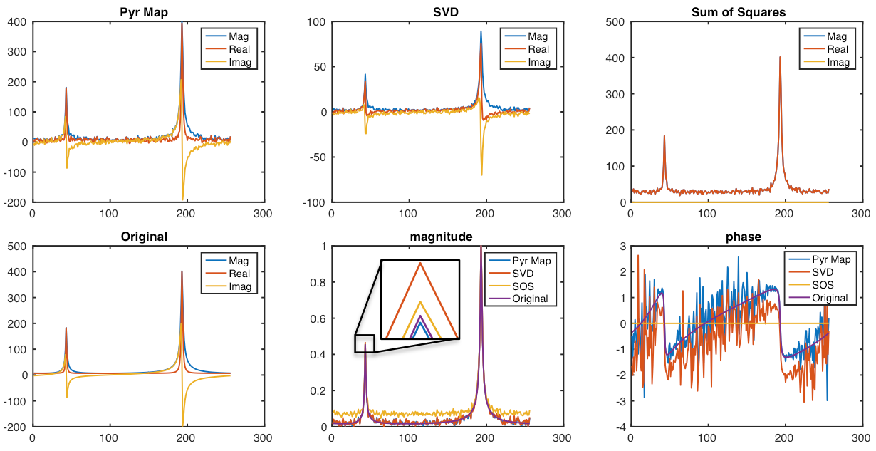
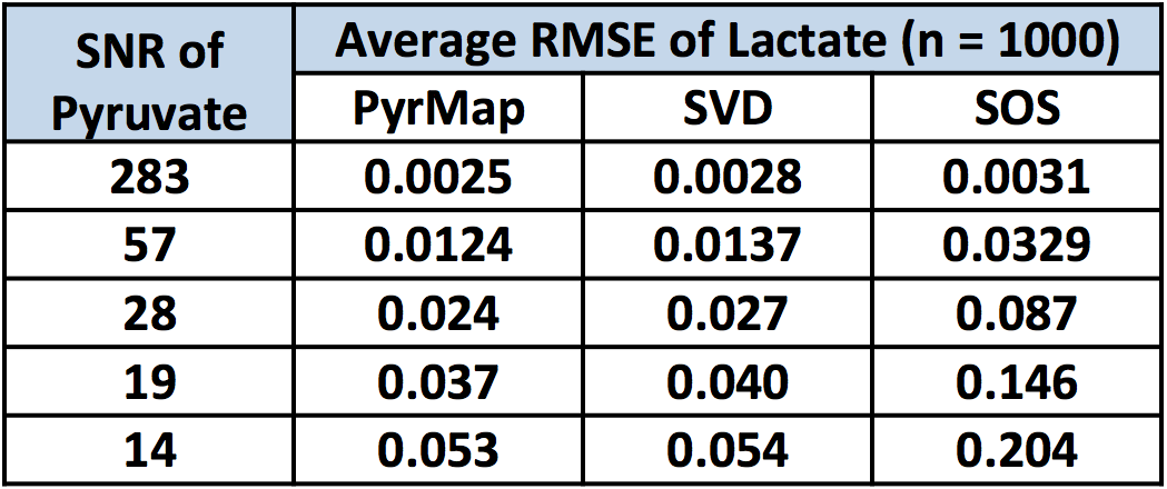
Figure 2 qualitatively demonstrated a representative simulation result, and table 1 quantitatively demonstrated the average RMSE of each reconstruction method at different noise levels compared to the ground truth. From Figure 2, at this noise level (SNR = 57), PyrMap method demonstrated the smallest RMSE compared to the ground truth as shown in the normalized magnitude image, and it also showed the most similar spectral phase compared to the ground truth. Simulation results in Table 1 demonstrated that both PyrMap and SVD methods performed more robustly than SOS method based on the RMSE.
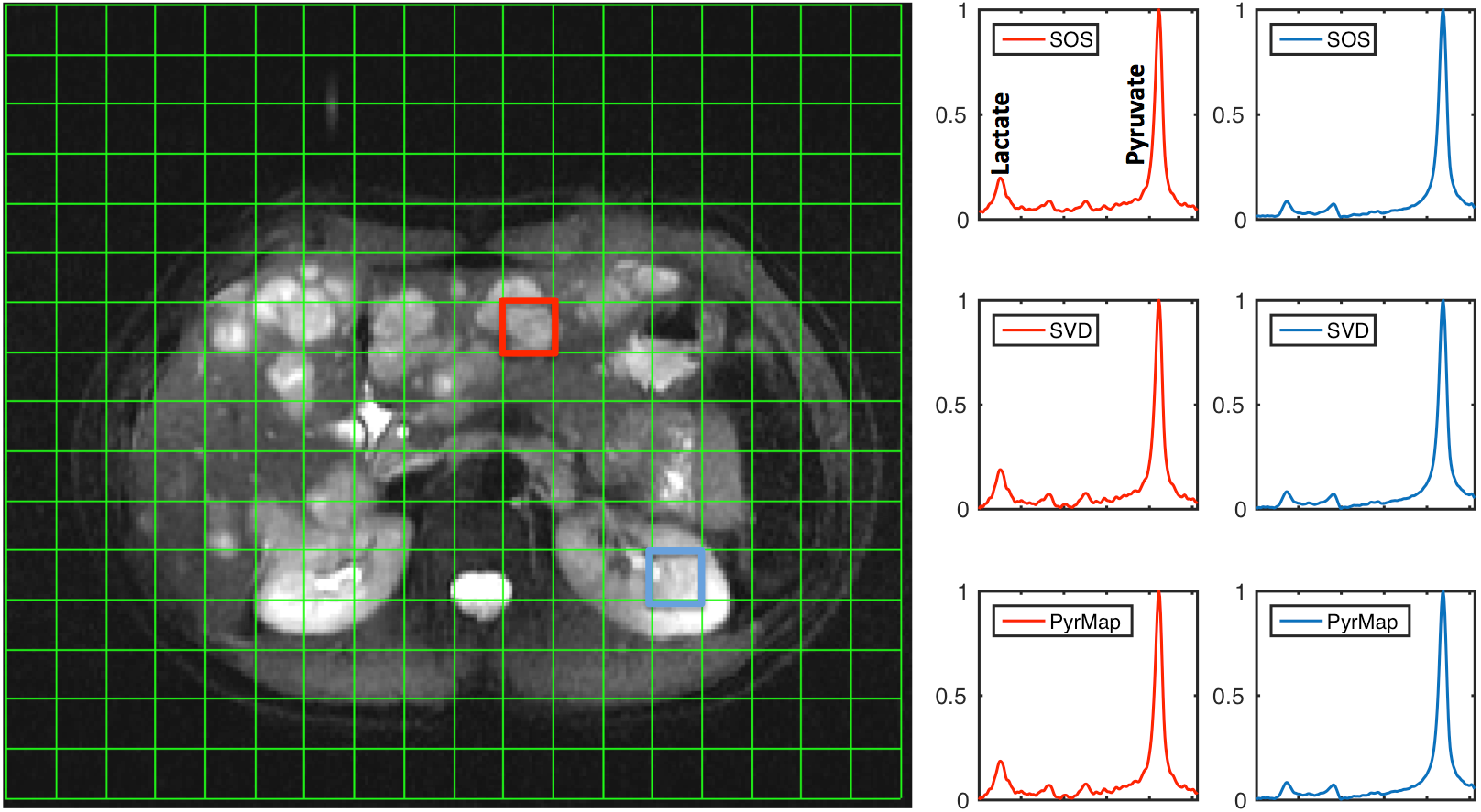
Figure 3 demonstrated the coil-combined magnitude spectra from selected voxels of a patient dataset. Overall, SOS showed an elevated baseline, while the PyrMap and SVD methods showed similar reconstructed spectra.
Discussion and Conclusions
This study demonstrated the acquisition and reconstruction of 16-channel hyperpolarized13C data throughout the liver and adjacent tissues in metastatic cancer patients. As shown by simulation in Table 1 and Figure 2, at various SNR levels, both PyrMap and SVD methods have demonstrated robust coil-combined results, while SOS generally provided a higher spectral baseline and was more prone to noise error. In addition, phase information was best preserved by PyrMap method, while SOS completely lost the phase information and SVD method introduced an arbitrary phase. This could potentially be problematic for other phase-sensitive acquisitions.
A spectral reconstruction framework was built to incorporate these methods for in vivo patient acquisitions using 16-channel coils. By qualitatively assessing the coil-combined data, PyrMap and SVD methods slightly outperformed the SOS method by preserving phase information and showing lower spectral baseline. Quantitative assessment was challenging in this case, due to lack of ground truth and different noise distributions (SOS noise was not Gaussian).
Overall, we have successfully developed a robust framework for multi-channel hyperpolarized 13C data combination and have demonstrated it in vivo. Extracting complex pyruvate signal provides an excellent approximation of the coil sensitivity map while maintaining valuable phase information of the coil-combined data.
Acknowledgements
This work was supported by NIH grants R01CA183071 and P41EB013598.References
[1] Nelson, Sarah J., et al. "Metabolic imaging of patients with prostate cancer using hyperpolarized [1-13C] pyruvate." Science translational medicine 5.198 (2013): 198ra108-198ra108.
[2] Rodgers, Christopher T., and Matthew D. Robson. "Receive array magnetic resonance spectroscopy: whitened singular value decomposition (WSVD) gives optimal Bayesian solution." Magnetic resonance in medicine 63.4 (2010): 881-891.
[3] Roemer, Peter B., et al. "The NMR phased array." Magnetic resonance in medicine 16.2 (1990): 192-225.
Figures