3829
Intravoxel incoherent motion (IVIM) imaging for differential diagnosing hepatocellular carcinoma (HCC), hepatic hemangioma(HHA) and hepatic metastasis(HM).1Radiology Department, The First Affiliated Hospital of Dalian Medical University, Dalian, China, 2GE Healthcare China, Beijing, China
Synopsis
The diffusion property of tumor tissues largely depends on cell density, which may also be predictive features of malignancy in some types of tumors. Intravoxel incoherent motion (IVIM) imaging is an extension of diffusion weighted imaging (DWI) that can be used to investigate both diffusion and perfusion changes in tissues. Comparing the IVIM parameters between carcinoma (HCC), hepatic hemangioma and hepatic metastasis, we found that IVIM can facilitates understanding of tumor tissue characteristics of perfusion and diffusion, and it may provide more useful information to distinguish hemangiomas from other two malignant tumors.
Purpose
Perfusion is an important phenomenon of many physiological or pathological processes [1]. Intravoxel incoherent motion (IVIM) imaging is an extension of diffusion weighted imaging (DWI) that can be used to investigate both diffusion and perfusion changes in tissues. It is reported that IVIM imaging may be useful for differentiating soft tissue tumors [2]. Our study was to assess the IVIM parameters for differential diagnosing hepatocellular carcinoma (HCC), hepatic hemangioma and hepatic metastasis.Methods
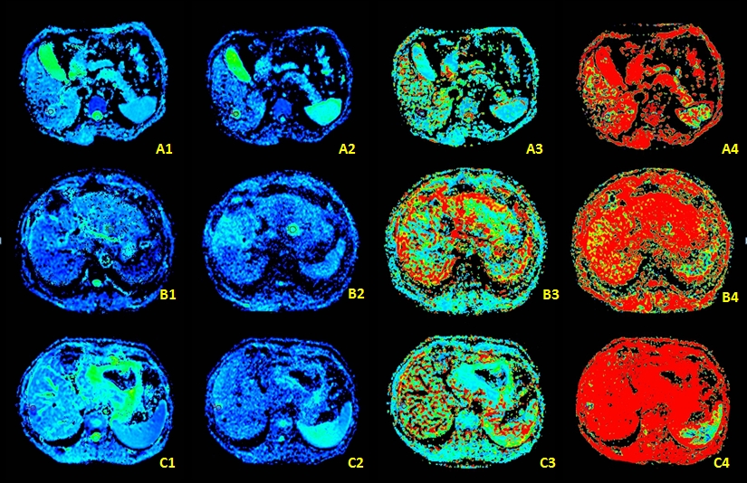
Seventy-one consecutive patients (40 men: 31 women, mean age, 62 years) with 33 hepatocellular carcinomas (HCCs), 24 hepatic hemangiomas and 14 hepatic metastasis were enrolled in this study and inspected conventional MR and IVIM-MR examination with 1.5-T MR imager from January 2015 to October 2015. MRI was performed using a 1.5-T MR imager (GE-Signa HDXT) in a protocol containing the routine T1WI, T2WI and 19 b values (0, 10, 30, 50, 80, 100, 120, 150, 180, 200, 300, 500, 800, 1000, 1200, 1500, 2000, 2500 and 3000 s/mm2) were used to evaluate diffusion and perfusion characteristics of IVIM. IVIM parameters (ADCstandard, ADCslow, ADCfast, and f) of hepatocellular carcinoma (HCC), hepatic hemangioma and hepatic metastasis were measured by using the FuncTool on GE AW4.6 workstation (Figure 1). The MR images were blindly reviewed and analyzed by two observers who have 3 and 10 years’ experience of MR diagnosis respectively. The SPSS17.0 statistical software has been used for the data analysis, P value less than 0.05 was considered statistically significant. Intraclass correlation coefficient (ICC) test was applied to test the consistency of two observers; compared ADCstandard, ADCslow, ADCfast and f of the three groups by Mann-Whitney test, respectively.Results and Discussion
The ICC values of the IVIM parameters (ADCstandard, ADCslow, ADCfast, and f) were all greater than 0.75 in the three groups, exhibiting an amenable consistency (Figure.2). The ADCstandard, ADCslow and f of the hepatic hemangioma were significantly higher (p < 0.05) than the hepatic metastasis (Figure.3). The ADCstandard, ADCslow and f of hepatic hemangioma were significantly higher (p < 0.05) than HCC, while the ADCfast of hepatic hemangioma were significantly lower (p < 0.05) than HCC (Figure.4).The ADCstandard, ADCslow, ADCfast and f did not show any statistically significant difference between the HCC and the hepatic metastasis (p > 0.05). IVIM MR imaging shows a unique profile of microcirculation and pure molecular diffusion within tumors. Our study showed that the ADCstandard values of hepatic hemangioma were significantly higher than hepatic metastasis (p < 0.05) and HCC (p < 0.05), since the limitation of water molecular diffusion of malignant tumors leads to a decrease of ADC value. The ADCslow value was another effective parameter to distinguish hemangiomas from the other two tumors. The ADCslow with a high b value is the true diffusion coefficient of pure water in tumors with perfusion components removed at the same time. For HCC and hepatic metastasis, the cell membrane permeability reduced, and the water molecular diffusion limited, so the ADCslow decreased significantly. The ADCfast of HCC was higher than hepatic hemangioma due to the affluent microvessel perfusion of HCC. The f value may correlate with the amount of normal angiogenesis with intact vessels in terms of basement membrane thickness and pericyte coverage, and it increases with the augmented tissue perfusion components. Our study showed that the f of hepatic hemangioma were significantly higher than hepatic metastasis (p < 0.05) and HCC (p < 0.05), because the hepatic hemangioma are rich in capillaries per unit tumor volume.Conclusion
According to our study, the HCC, hepatic metastasis and hepatic hemangioma showed different IVIM parameters (ADCstandard, ADCslow, ADCfast and f values). IVIM imaging facilitates understanding of tumor tissue characteristics of perfusion and diffusion, and it may provide more useful information to distinguish hemangiomas from other two malignant tumors.Acknowledgements
No acknowledgement found.References
1.Le B D. Intravoxel incoherent motion perfusion MR imaging: a wake-up call.[J]. Radiology, 2008, 249(3):748-52.
2.Penner AH, Sprinkart AM, Kukuk GM, et al. Intravoxel incoherent motion model-based liver lesion characterisation from threeb-value diffusion-weighted MRI. EurRadiol, 2013;23:2773–2783.
3.Wagner M, Doblas S, Daire JL, et al. Diffusion-weighted MRimaging for the regional characterization of liver tumors. Radiology 2012;264:464–472.
4.Melanie Chiaradia M D, Laurence Baranes M D, Nhieu J T V, et al. Intravoxelincoherent motion (IVIM) MR imaging of colorectal liver metastases: Are we only looking at tumor necrosis?[J]. Journal of Magnetic Resonance Imaging Jmri, 2014, 39(2):317-325.
5.Chow A M, Gao D S, Fan S J, et al. Liver fibrosis: An intravoxel incoherent motion (IVIM) study[J]. Journal of Magnetic Resonance Imaging Jmri, 2012, 36(1):159-167.
6.Suo S, Cao M, Zhu W, et al. Stroke assessment with intravoxel incoherent motion diffusion-weighted MRI[J]. Nmr in Biomedicine, 2016, 29(3):320-328.
Figures