3817
The Restricted Diffusion Model for Differentiation of Tumor from Normal Tissue in Glioblastoma1Biomedical Engineering, University of Michigan, Ann Arbor, MI, United States, 2Radiation Oncology, University of Michigan, Ann Arbor, MI, United States, 3Radiology, University of Michigan, Ann Arbor, MI, United States
Synopsis
It is a challenge to differentiate non-enhanced solid tumor from edema in glioblastoma. This study applied the restricted diffusion model to high b-value diffusion weighted images to characterize glioblastoma. The formation of the restricted diffusion model was derived for bi-polar diffusion gradients. The parameters fitted by the restricted diffusion model can differentiate solid tumor from edema and normal-appearing white matter and grey matter, better than the conventional apparent diffusion coefficient and the bi-exponential model without accounting for diffusion restriction of intra-cellular water.
Introduction
Restricted water diffusion in glioblastoma (GBM) has been reported. However, the conventional apparent water diffusion (ADC) is often elevated and cannot differentiate solid tumor from edema in GBM.1-5 A recent study shows that cell sizes and cellularity in three colon cancer cell lines estimated by a restricted diffusion model (RDM) were correlated with histology.6 The RDM considers intra-cellular water diffusion restricted in spherical cells and modulated by diffusion gradients,6-8 and its formulations are derived for the mono-polar pulse gradient spin echo and oscillating gradient spin echo.6-7,9
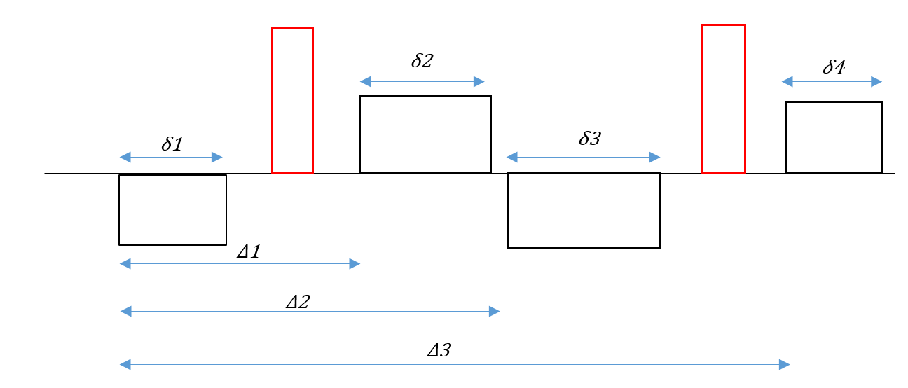
In this study, we extended the RDM formulation to a bi-polar pulse gradient spin echo (Figure 1) (that minimizes eddy-current caused artifacts in diffusion images). We characterized GBM using this model and compared to normal-appearing white matter (WM) and grey matte (GM), and edema.
Methods and Materials
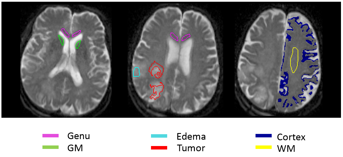
Using the Gaussian phase approximation, the intracellular diffusion signal restricted in spheres and modulated by bi-polar diffusion gradients can be expressed as7,9: $$S_{in}=\frac{\gamma ^{2}}{2}\sum B_{n}\int_{0}^{2\epsilon}dt_{1}g(t_{1})\int_{0}^{2\epsilon}g(t_{2})exp(-D_{in})\lambda_{n}\left | t_{1}-t_{2} \right |dt_{2}$$where Din is an intracellular diffusion coefficient, λn and Bn are structure dependent parameters related to the spherical cell radius R ,7,9 and g(t) represents diffusion gradient waveforms. For the bi-polar diffusion gradients in Figure 1, we have:$$\begin{cases} g& \text{ if } \ 0<x<\delta _{1} \\ g& \text{ if } \Delta _{1}<x<\Delta_{1}+\delta_{2} \\ -g& \text{ if } \Delta _{2}<x<\Delta_{2}+\delta_{3} \\ -g & \text{ if } \Delta _{3}<x<\Delta_{3}+\delta_{4} \end{cases}$$The integral in first equation (that is not shown here) is derived for the intra-cellular diffusion signal. The extracellular diffusion signal can be expressed as:$$S_{ex}=exp(-bD_{ex})$$ where Dex is extra-cellular diffusion coefficient. The total water diffusion signals can be depicted as: $$$S=V_{in}S_{in}+(1-V_{in})S_{ex}$$$, where Vin is the fractional volume of intra-cellular water.Thirty patients with newly histologically diagnosed GBM were studied. Diffusion weighted images (DWIs) were acquired with 11 b values from 0 to 2500 s/mm2 on a 3T scanner (Skyra, Siemens Healthineers). DW signals in volumes of interest (VOIs) (Figure 2) of solid tumor (determined from a previous study), edema (distance from tumor and recurrence but with T2 elevation), and 2 white matter (WM) and 2 grey matter (GM) were fitted to the RDM to estimate parameters of R, Din, Dex and Vin using simplex algorithm in Matlab. Fitting was run multiple times, and the results with the minimum MSE were accepted. Whether the fitted parameters could differentiate solid tumor from other tissue types was evaluated by Students’ t-test. For comparison, the conventional apparent diffusion coefficient (ADC) obtained from a mono-exponential fitting of DWIs with b-values from 0-1000 s/mm2 was evaluated for distinguishing solid tumor from other tissue types. A similar evaluation was performed for the parameters (2 diffusion coefficients (D1 and D2) and the fractional volume (Vin)) from the conventional 2-exponential model.Results
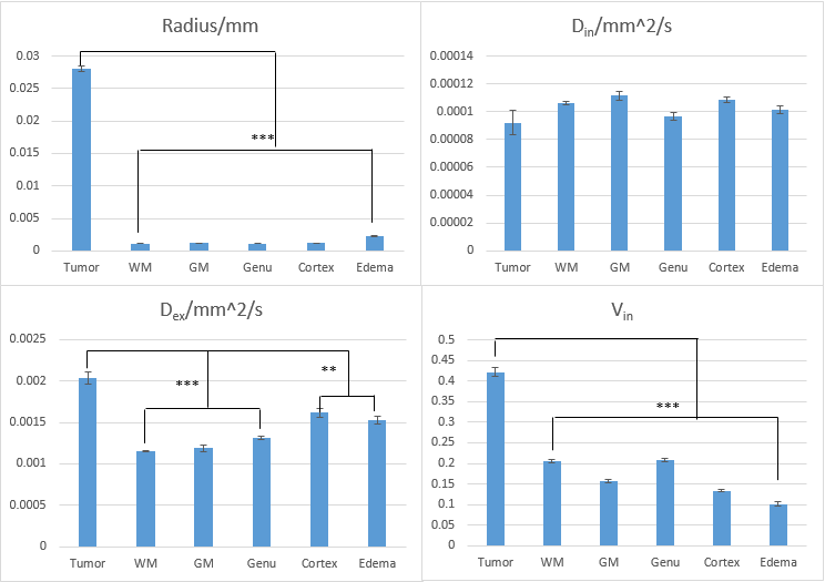
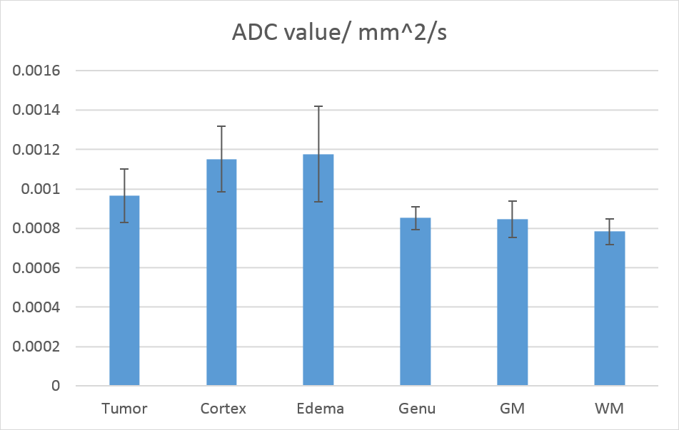
R, Dex and Vin derived from the RDM in tumor VOIs were significantly greater than ones in frontal WM, genu, head of caudate nucleus, cortex and edema (p < 0.01-0.001), but not Din (Figure 3). Most interesting, R values in solid tumors, ranged from 21.6 to 34.5 um, did not have any overlap with ones (from 0.9 to 3.5 um) in all other tissue types. Similarly, Vin values in solid tumors, ranged from 0.32 to 0.52, again did not overlap with the values (from 0.05 to 0.25) in all other tissue types. Elevated Dex in cortex compared to deep GM could be primarily due to the partial volume effect of cerebral spinal fluid. In the conventional 2-exponential model, none of the three parameters could significantly differentiate tumor from all other tissue types (Figure 4). D1 in tumor (2. 02±0. 07 um2/ms) was significantly different from ones in WM and GM but not from edema (1. 89±0. 06 um2/ms). Also, D2 in solid tumor (0. 34±0. 01 um2/ms) was not significantly different from deep GM (0. 36±0. 02 um2/ms) and edema (0. 33±0. 04 um2/ms). Vin of tumor (0. 42±0. 01) was not significantly different from frontal WM (0. 38±0. 01) and deep GM (0. 46±0. 02). The conventional ADC could not significantly differentiate tumor from any other tissue types (Figure 5), which is similar to other reports.1-5
Discussion and Conclusion
In this study, we fitted the restricted diffusion model to DWIs acquired with bi-polar diffusion gradients in glioblastoma. The explicit consideration of intra-cellular water diffusion restriction in the model can differentiate tumor from other tissue types better than without, particularly from edema. The tumor VOI used in this study was correlated with progression free survival. Further investigation will be carried out by combining with CBV for better definition of tumor volume and prediction of therapy outcome in GBM.Acknowledgements
This work is supported in part by a grant of NIH/NCI 1U01CA183848.
Thanks for Dr. Himanshu Bhat (Siemens Healthineers) for assistance of the project.
References
1. P. Pramanik, H. Parmar, A. Mammoser, L. Junck, M. Kim, C. Tsien, T. Lawrence and Y. Cao, "Hypercellularity Components of Glioblastoma Identified by High b-Value Diffusion-Weighted Imaging", International Journal of Radiation Oncology*Biology*Physics, vol. 92, no. 4, pp. 811-819, 2015.
2. T. Sugahara, Y. Korogi, M. Kochi, I. Ikushima, Y. Shigematu, T. Hirai, T. Okuda, L. Liang, Y. Ge, Y. Komohara, Y. Ushio and M. Takahashi, "Usefulness of diffusion-weighted MRI with echo-planar technique in the evaluation of cellularity in gliomas", Journal of Magnetic Resonance Imaging, vol. 9, no. 1, pp. 53-60, 1999.
3. T. Chenevert, "Diffusion Magnetic Resonance Imaging: An Early Surrogate Marker of Therapeutic Efficacy in Brain Tumors", Journal of the National Cancer Institute, vol. 92, no. 24, pp. 2029-2036, 2000.
4. H. Lyng, O. Haraldseth and E. Rofstad, "Measurement of cell density and necrotic fraction in human melanoma xenografts by diffusion weighted magnetic resonance imaging", Magnetic Resonance in Medicine, vol. 43, no. 6, pp. 828-836, 2000.
5. A. Guo, T. Cummings, R. Dash and J. Provenzale, "Lymphomas and High-Grade Astrocytomas: Comparison of Water Diffusibility and Histologic Characteristics", Radiology, vol. 224, no. 1, pp. 177-183, 2002.
6. X. Jiang, H. Li, J. Xie, E. McKinley, P. Zhao, J. Gore and J. Xu, "In vivo imaging of cancer cell size and cellularity using temporal diffusion spectroscopy", Magnetic Resonance in Medicine, vol. 78, no. 1, pp. 156-164, 2016.
7. J. Xu, M. Does and J. Gore, "Quantitative characterization of tissue microstructure with temporal diffusion spectroscopy", Journal of Magnetic Resonance, vol. 200, no. 2, pp. 189-197, 2009.
8. A. Ianuş, B. Siow, I. Drobnjak, H. Zhang and D. Alexander, "Gaussian phase distribution approximations for oscillating gradient spin echo diffusion MRI", Journal of Magnetic Resonance, vol. 227, pp. 25-34, 2013.
9. J. Stepišnik, "Time-dependent self-diffusion by NMR spin-echo", Physica B: Condensed Matter, vol. 183, no. 4, pp. 343-350, 1993.
Figures