3806
Use of a Rigidity Penalty to Improve MR-CT Image RegistrationElizabeth MaryAnn McKenzie1, Dan Ruan2, Percy Lee2, and Ke Sheng2
1Physics and Biology in Medicine, University of California Los Angeles, LOS ANGELES, CA, United States, 2Radiation Oncology, University of California Los Angeles, Los Angeles, CA, United States
Synopsis
Attenuation coefficients of tissue must be known to accurately model dose in radiation therapy. MR-guided radiation therapy better visualizes soft tissue, but it does not inherently contain attenuation information in the same way as CT images. CT and MR images can be registered to pool information, but the bones can become severely distorted. This work applies a rigidity penalty to bones segmented in CT, such that the soft tissue is allowed to deform while the bones remain rigid during CT to MRI registration. We show that this technique improves the registration, making the deformations more anatomically feasible.
Introduction
MRI-CT registration is commonly used in MR-based radiation treatment planning and delivery. While MRI provides superior soft tissue contrast, CT values can be directly used to calculate radiation attenuation and dose. However, the multimodality registration between MRI and CT often results in distortion of the bony anatomy, which may have undesired dosimetric consequences (Figure 1). One cause of this distortion is the difficulty in visualizing bone in MRI relative to CT. This study aims to improve multimodality registration accuracy by including bone rigidity regularization in the deformable registration cost function. Improving MRI-CT registration will aid in MR-based radiation therapy, as well as aid those who wish to fully utilize the complimentary information contained within MRI and CT.Methods
Nine patients with both CT and MR images were selected for this study. The bones were first segmented on the CT images using thresholding. A B-spline based deformable registration was performed to maximize mutual information of the deformed CT and target MR images while preserving the rigidity of the segmented bone1. This was done using a rigidity penalty2, which enforces affinity, orthonormality, and properness in labeled rigid regions. While this method was initially developed to address CT-CT registration, MR-CT registration is more challenging due to inherent differences in contrast. For the first time, we applied this technique in the setting of multi-modality registration. The bone rigidity regularized registrations using the proposed method are compared to rigid registrations, and B-spline deformable image registrations without the rigidity penalty. This is done for three sites: the brain, abdomen and pelvis. In addition to visual inspection of the registration quality, the determinant of the spatial Jacobian was used to evaluate the preservation of the bone volumes.Results
Visually, rigidity-regularized deformable registration matches the exterior soft tissue better than the rigid registration, while maintaining better bone spatial integrity compared to the B-spline deformable registration (Figure 2). Using the rigidity penalty in deformable registration significantly reduced the average maximum of the Jacobian determinant from 20.9 to 2.04 across the entire imaging volume, indicating an ability to control large, unfeasible deformations (Figure 3). Within the bone volumes, the average Jacobian determinant was improved from 0.844±0.543 to 0.974±0.0574 (Figure 4). A value of unity is ideal (indicating no compression or decompression). The improved bone volume preservation was not site dependent. Additionally, the use of the bone rigidity regularizer reduced the incidence of anatomically unfeasible folds in the image space, as shown by the reduction in negative Jacobian determinants (Figure 4, “Min” columns).Discussion
We demonstrated a novel MRI-CT registration method using a rigidity penalty on bone while allowing soft tissues to deform. This leads to an improved balance in the flexibility of deformable registration as well as a higher degree of anatomical fidelity. Since bony anatomy can have some of the largest effects on attenuation and dose calculation3, correcting for these distortions is an important consideration in MR-guided radiation therapy treatment planning. For the purpose of radiotherapy, an important benefit of this technique lies in the similar voxel intensity between bone and air in MR images4. This similarity can challenge an image registration algorithm, and it may mistakenly stretch the bones into nearby volumes with low electron density such as the lung and sinuses (or vice versa) if the bone rigidity is not preserved. Bone and lung have drastically different attenuation coefficients, and replacing one with the other could lead to considerable errors in calculated dose. The proposed technique regulates the allowable motion of the bones, and thus prevents this kind of mismatch when using CT-derived attenuation coefficients in an MR planning image. While the purely rigid registration also prevents these types of errors, the bony anatomy is not allowed to flex, and the soft tissue also remains unrealistically rigid.Conclusion
Including a bone rigidity term in the deformable registration significantly improves MRI-CT registration and reduces the confusion of bone-air volumes, which is particularly important for MR-based dose calculation accuracy. Compared to rigid registration, this new framework allows greater freedom in matching soft tissues.Acknowledgements
I'd like to thank my adviser Dr. Sheng for all of his support and encouragement. I'd also like to thank the people who generously share their source code and ideas online. They make so much of today's research possible.References
- Klein S, Staring M, Murphy K, Viergever MA, Pluim JPW. elastix : A Toolbox for Intensity-Based Medical Image Registration. 2010;29(1):196-205.
- Staring M, Klein S, Pluim JPW. A rigidity penalty term for nonrigid registration. Med Phys. 2007;34(11):4098-4108.
- Uh J, Merchant TE, Li Y, Li X, Hua C. MRI-based treatment planning with pseudo CT generated through atlas registration. Med Phys. 2014;41(5):51711.
- Schreibmann E, Nye JA, Schuster DM, Martin DR, Votaw J, Fox T. MR-based attenuation correction for hybrid PET-MR brain imaging systems using deformable image registration. Med Phys. 2010;37(5):2101-2109.
Figures
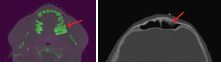
Example of CT to MRI
registration in a head and neck case using a B-spline model without a rigidity
constraint. The severe distortion in the
molars is quite apparent in the left image.
In the right image, the bone has been distorted into the air cavity.
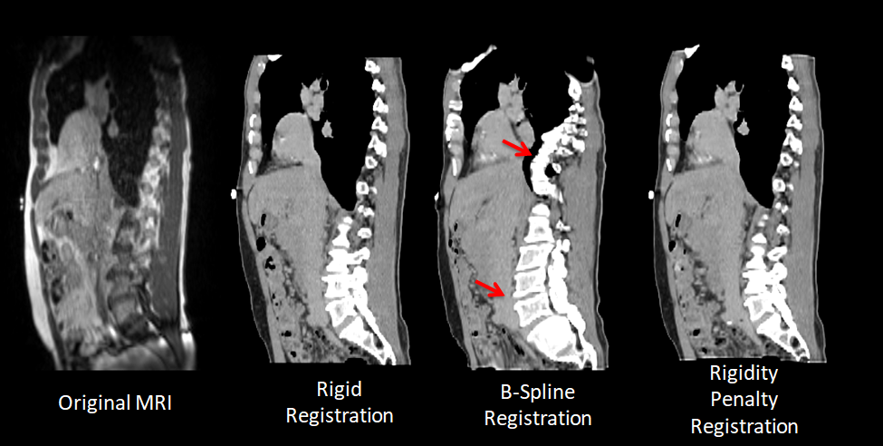
Example of
registration results compared to the fixed MR image (far left). The Rigid Registration shows adequate
alignment, however the soft tissue has not been deformed to match the MRI. While the soft tissue has deformed to better
match the MRI, the bones are only rigidly moved in the Rigidity Penalty
Registration. This can be seen by the non-rigid
deformation of the heart while the spinal column remained intact. The B-spline registration shows anatomically
unfeasible deformation of the spinal column, as indicated by the arrows, while
the anterior portion of the heart has deformed to match the MRI.
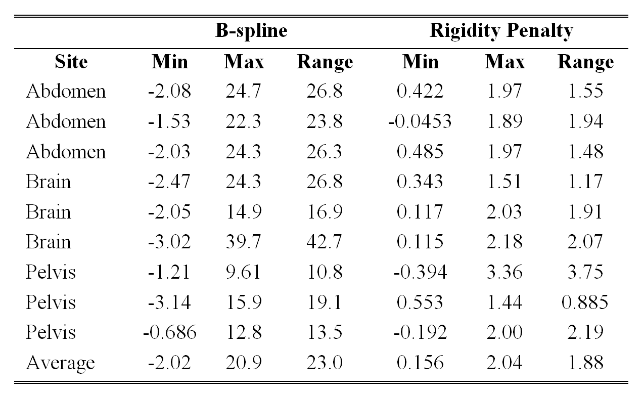
Jacobian Determinant
for both B-spline and Rigidity Penalty registrations across the entire image
for each image.
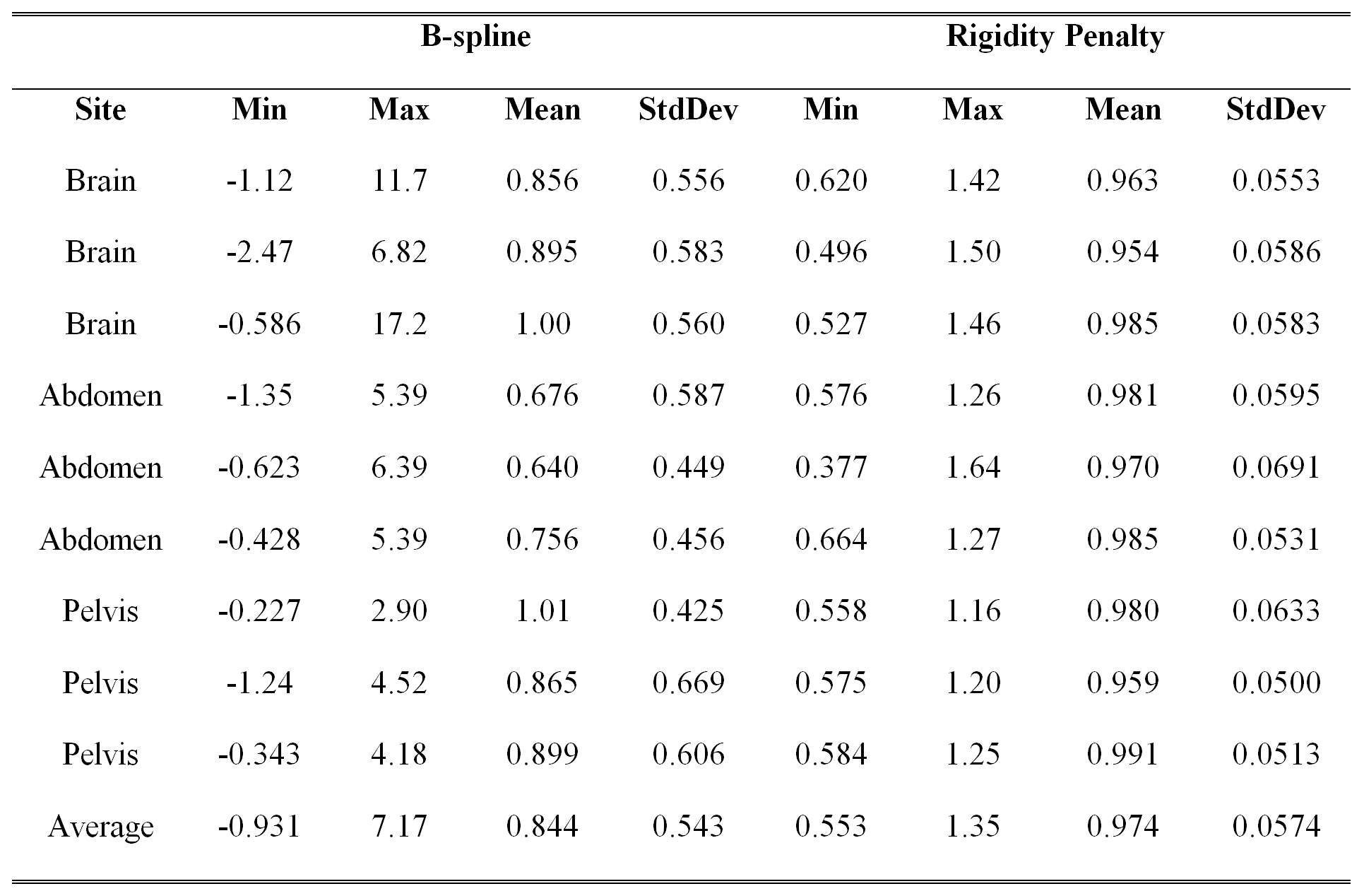
Statistics on
Jacobian determinant voxels contained within the rigidity penalty region.