3790
Multi-modal MRI investigation of brain plasticity during prolonged Braille learning in sighted subjects – voxel-based morphometry and quantitative T1 mapping approach1Laboratory of Brain Imaging, Nencki Institute of Experimental Biology, Warsaw, Poland, 2Department of Psychology, Jagiellonian University, Kraków, Poland, 3Academy of Special Education, Warsaw, Poland, 4Laboratory of Psychophisiology, Nencki Institute of Experimental Biology, Warsaw, Poland
Synopsis
In order to study structural brain reorganization, multi-modal MRI combining voxel-based morphometry and quantitative T1 mapping was used in longitudinal design on sighted subjects who underwent tactile Braille reading course. Results show that methods are complimentary to each other. Combined approach like that gives insight into different aspects of tissue property changes introducing new opportunities for studying brain plasticity.
Introduction
Multi-modal magnetic resonance imaging (MRI) refers to many diverse ways of effectively combining data from various MRI contrasts. Voxel-based morphometry (VBM) is the dominant method of analyzing non-quantitative T1-weighted (T1w) high-resolution structural magnetic resonance images. It has been widely used to study gray matter volume (GMV) reorganization due to disease progression or training of new skill1,2. Relatively novel method of quantitative T1-mapping (qT1m) provides signal sensitive to gray matter myelination3,4. By definition brain plasticity is driven mainly by a mismatch between functional supply and environmental demand5. One of the tasks that is often used to investigate large-scale neuroplasticity in the brain of both blind and sighted subjects is Braille reading6,7,8. Here for the first time we applied both VBM and qT1m in longitudinal brain plasticity study on sighted subjects who underwent tactile Braille reading course. The employed multi-modal approach can inform of different aspects of tissue property changes introducing new opportunities for studying brain plasticity.Methods
28 right–handed females (Age M = 23.3; SD = 3.5) underwent tactile Braille reading course lasting 8 months with 5 MRI sessions. After first 3 months 14 subjects decided to read with left hand. MRI sessions were conducted every 3 months: before the course (TP0), 3 during tactile reading acquisition (TP1, TP2, TP3) and 3 months after the end of the course (TP4).
Data were collected on 3T Siemens scanner with 12-channel coil using standard structural T1-weighted MPRAGE sequence as well as T1-mapping by Variable Flip Angle method. For VFA T1-map we used 3D gradient-echo, 1mm-isotropic resolution scans with FA=4 and FA=18 deg (TE=2.46 ms, TR=7.6ms, TA=2*5min27s). Maps were corrected for flip angle error by B1+ map (stimulated echo to spin echo ratio)6.
Analysis of MRI data was performed using MATLAB environment in SPM12, CAT12 and house-build scripts for qT1m reconstruction. For each participant, the T1w scans from all time points were registered and bias-corrected using the Serial Longitudinal Registration in SPM12, which combines diffeomorphic and rigid-body registration. The output consisted of an average T1w image and five within-subject Jacobian difference maps was a base for the further analyses. Then each participant's average image was segmented to obtain native space tissue probability maps and DARTEL normalization input. For VBM within-subject analyses we used a product of Jacobian difference at each time point and GM TPM normalized with the DARTEL template. The average T1w was segmented in CAT12 with an option of cortical surface reconstruction. Each reconstructed qT1m was then sampled with the native space central cortical surface resulting in five identical surfaces false coloured with adjacent T1 relaxation values for each subject. All the data was smoothed voxel- or surface-wise and analyzed in mixed-ANOVA with subject, time and hand preference as factors.
Results
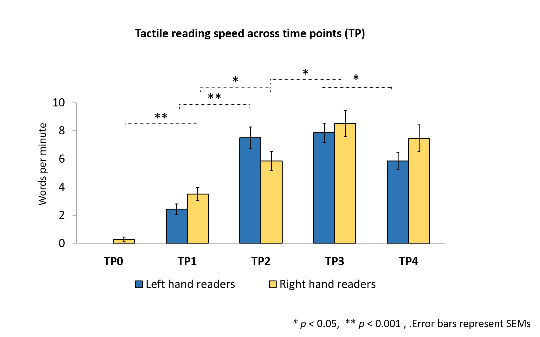
Subjects successfully acquired tactile Braille reading, improving their reading speed over time. There was no effect of preferred hand, nor time by hand interaction (Figure 1).
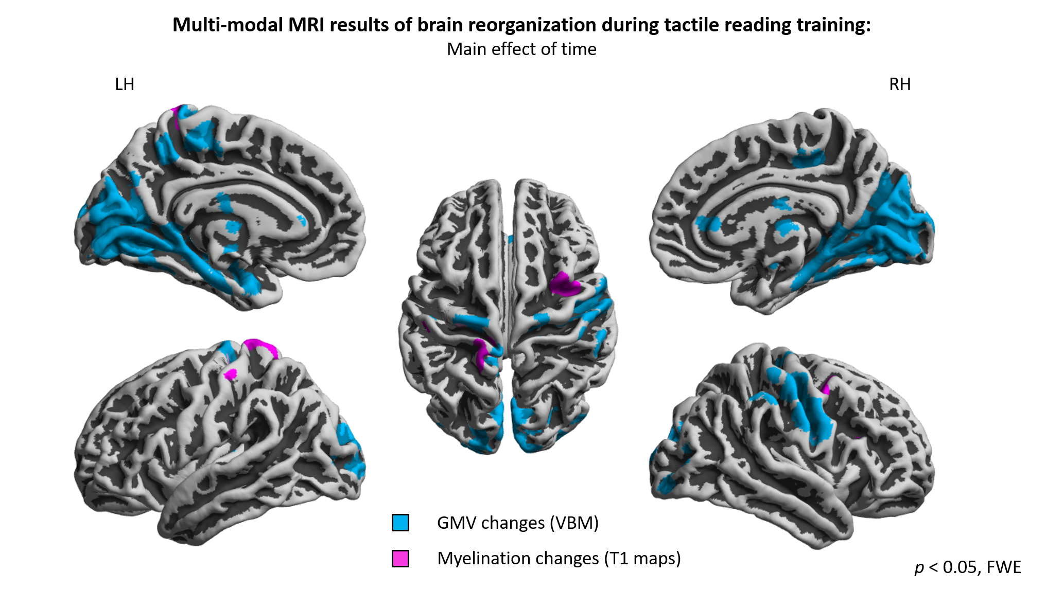
In VBM analysis significant main effect of time point showed that tactile reading training induced GMV changes in various brain regions including visual, premotor, motor and somatosensory cortices (Figure 2). Post-hoc analysis showed that GMV increase in calcarine and cuneal cortex occurs both in right- and left-hand readers, while motor and somatosensory reorganization is specific to left-hand readers.
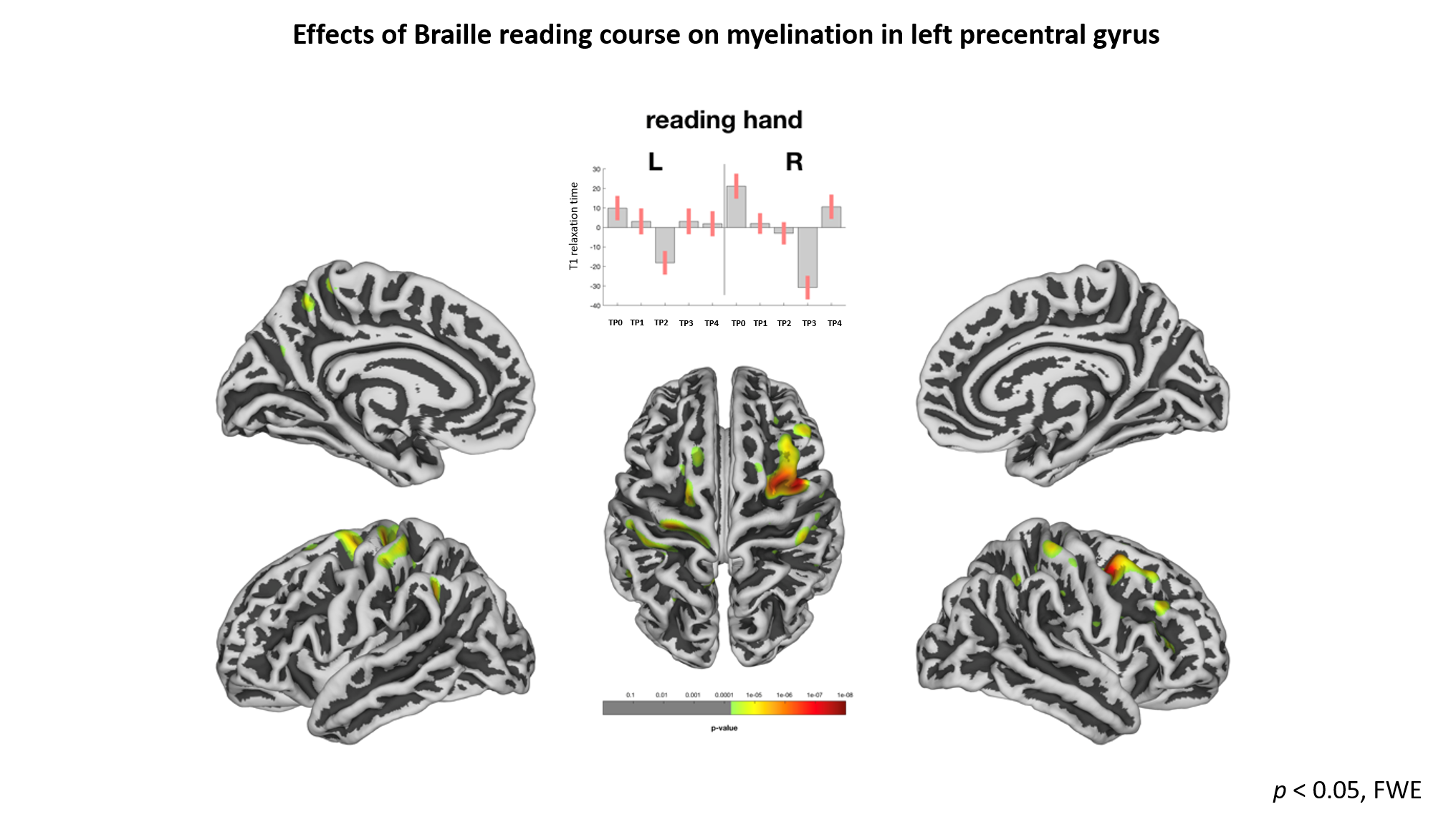
In qT1maps analysis significant main effect of time showed
regions in left pre- and central gyrus and right middle frontal gyrus. Dynamics
of the changes follows the progress of Braille learning curve in the group of
right-hand readers (Figure 3).
Conclusions
Our longitudinal study revealed large-scale brain reorganization due to tactile reading training in sighted subjects. Structural and quantitative methods provide complementary information about gray matter reorganization, showing regions associated with better Braille reading. What is interesting gray matter volume changed in right hemisphere motor cortex, while gray matter myelination in left pre- postcentral gyrus. Nevertheless, lateralization in both methods was specific to reading hand. Reorganization in the visual cortex was observed only in VBM, which shows that combining different MRI methods may lead to broader understanding of neural processes underlying brain plasticity. We plan to compare results to control group in order to investigate specificity of effects to tactile reading training.Acknowledgements
This project was realized with the aid of CePT research infrastructure purchased with funds from the European Regional Development Fund as part of the Innovative Economy Operational Programme, 2007–2013. Study was conducted with National Science Center grant Harmonia UMO-2014/14/M/HS6/00918.References
1. Draganski, B., Gaser, C., Busch, V., Schuierer, G., Bogdahn, U., May, A., 2004. Neuroplasticity: changes in grey matter induced by training. Nature 427, 311–312.
2. Woollett, K., Maguire, E.A., 2011. Acquiring “the knowledge” of London's layout drives structural brain changes. Curr. Biol. 21, 2109–2114.
3. Dick, F., Tierney, A.T., Lutti, A., Josephs, O., Sereno, M.I., Weiskopf, N., 2012. In vivo functional and myeloarchitectonic mapping of human primary auditory areas. J Neurosci.32(46): 16095–16105.
4. Sereno, M.I., Lutti, A., Weiskopf, N., Dick, F., 2013. Mapping the human cortical surface by combining quantitative T(1) with retinotopy. Cereb Cortex. (9):2261-8
5. Lovden,M., Backman, L., Lindenberger, U., Schaefer, S., Schmiedek, F., 2010. A theoretical framework for the study of adult cognitive plasticity. Psychol. Bull. 136, 659–676
6. Büchel C, Price C, Friston K. 1998. A multimodal language region in the ventral visual pathway. Nature 394:274– 277. doi: 10.1038/28389
7. Reich L, Szwed M, Cohen L, Amedi A. 2011. A ventral visual stream reading center independent of visual experience. Current Biology:CB 21:363–368. doi: 10.1016/j.cub.2011.01.040
8. Siuda-Krzywicka, K., Bola, L., Paplińska, M., Sumera, E., Jednoróg, K., Marchewka, A., Sliwinska M., Amedi A., Szwed M. 2016. Massive cortical reorganization in sighted Braille readers. Elife 5:e10762. doi: 10.7554/eLife.10762
9. Preibisch C, Deichmann R., 2009. Influence of RF spoiling on the stability and accuracy of T1 mapping based on spoiled FLASH with varying flip angles. Magn Reson Med.(1):125-35
Figures