3789
Motion of the cerebellum in patients with Chiari malformation compared to healthy subjects: A quantitative study using spiral cine DENSE MRI1Radiology and Imaging Sciences, Emory University, Atlanta, GA, United States, 2Mechanical Engineering, University of Akron, Akron, OH, United States, 3Research and Development, Siemens Healthineers, Atlanta, GA, United States
Synopsis
The goal of this study was to acquire mid-sagittal cine DENSE images to quantify displacement and strain over the cardiac cycle in patients with Chiari malformation and age-matched controls. The major finding of this study was that both tissue displacement and tissue strain in the parenchyma of the cerebellum was significantly greater in patients with Chiari malformation than healthy controls.
Introduction
Brain tissue undergoes pulsatile motion due to cardiac-driven pressure/flow variations over each heartbeat. Altered brain motion has been documented in certain pathologies, including intracranial brain tumors and Chiari malformation (1,2). Recently, cardiac-gated, cine, balanced steady state free procession (bSSFP) has been used to examine cerebellum motion in subjects with Chiari malformation (3). Measurement and examination of cerebellar tonsillar motion using conventional bSSFP cine imaging is limited to tracking pre-defined anatomical landmarks over the cardiac cycle and cannot quantify displacement information over the brain parenchyma. Therefore, bSSFP is unable to quantify tissue strain which has been shown to be important in cellular signaling and tissue remodeling. Spiral cine imaging coupled with a Displacement Encoding using Stimulated Echoes (DENSE) sequence offers a method to quantify very small levels of brain tissue displacement on a pixel-by-pixel basis over the entire imaging slice (4,5). The goal of this study was to acquire mid-sagittal cine DENSE images to quantify displacement and strain over the cardiac cycle in patients with Chiari malformation and age-matched controls. We hypothesized that peak displacement and peak strain in the cerebellum and cerebellar tonsils would be higher in patients with Chiari malformation than in controls.Methods
We examined 9 adult healthy volunteers (age 28+/-4) and 12 patients (age range 31+/-6) with Chiari malformation. The Chiari diagnosis was confirmed by previous clinically-ordered imaging study (> 5m mm of tonsillar decent below the foreman magnum) and by clinical symptoms. All patients were referred by a single neurosurgeon (DB) and were under consideration for decompression surgery. All patients gave informed consent for a separate imaging study that included a cardiac-gated, spiral cine DENSE sequence performed at the mid-sagittal plane on a 3T MRI scanner (Siemens Prisma, Erlangen, Germany). Acquisition parameter included: two directions of motion encoding, encoding frequency, ke = 0.6-1.2 cycles/mm, two spiral interleaves per heartbeat, pixel size = 1.2 × 1.2 mm2, slice thickness = 7 mm, averages = 4, frames = 16-27, depending on the heart rate. Using an in-house written MATLAB program (DENSEpro), regions of interest (ROIs) were traced in multiple brain regions (including the cerebellum) on the magnitude images and copied to the phase images, figure 1. For each ROI on each participant, displacement magnitudes were spatially averaged for each cardiac frame. The frame with the largest average displacement was defined as the peak displacement in each subject. Principal strain was calculated using a structured quadrilateral grid on the two-dimensional displacement field. Peak principal strain was determined for each subject in same manner outlined for peak displacement. Peak displacement and peak strain were compared between the healthy controls and the Chiari patients using a student’s t-test. Spatial variation of tissue displacement over various brain regions was assessed using Lagrangian pixel-wise trajectories (6).Results
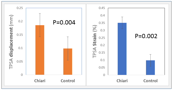
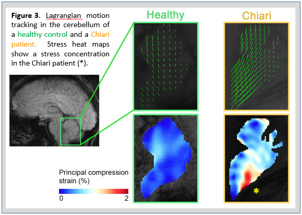
Spiral cine DENSE images were successfully acquired in all volunteers and all Chiari patients. Peak tissue displacements in the cerebellum were significantly higher in the Chiari patients than in the healthy controls (0.19 ± 0.08 mm vs 0.09 ± 0.04 mm, p=0.004). Similarly, Peak principal strain values in the cerebellum were significantly higher in the cerebellum in the Chiari patient compared to the health subjects (0.77% ± 0.33% vs 0.35% ± 0.10%, p=0.002), figure 2. Spatial variation of displacement and principal strains with the cerebellum were assessed using pixel-wise trajectories and strain maps, respectively. Figure 3 shows this analysis for a representative healthy control and a Chiari patient. The cerebellar tonsils had noticeably more anterior-posterior motion compared to more superior tissues. The spatial gradient of displacement resulted in localized elevated strain values within the cerebellum in both healthy and patient cases, but strain values were markedly larger in Chiari patients than in controls.Discussion
The major finding of this study was that displacement and strain of brain parenchyma in the cerebellum was significantly greater in patients with Chiari malformation than in healthy controls. The increased strain value was often characterized by localized increase in strain values in the inferior/posterior regions of the cerebellum. This finding is noteworthy as this sub-occipital anatomical region is often cited by Chiari patients as a region of pain during coughing, sneezing, or laughing. Further studies will examine temporal characteristics of motion, the motion and strain in other brain regions, and examine motion patterns in patients after decompression surgery.Conclusion
We have demonstrated that cardiac-gated, spiral cine DENSE imaging can measure spatially and temporally detailed variation of motion patterns in the brain and these motion patterns can be used to determine tissue strain values. In patients with Chiari malformation, both strain and motion were greater than in healthy controls.Acknowledgements
No acknowledgement found.References
1. Hofmann, E., et al. Amer J of Neuroradiology, 2000. 21(1): p. 151-158
2. Leung V, et al. J Neurosurg Spine. 2016 Apr;24(4):546-55.
3. Radmanesh A, et al. Neuroradiology, 2015 Apr;57(4):387-93.
4. Soellinger, M., et al. Magn Reson Med, 2009. 61(1): p. 153-62.
5. Zhong, X., et al. Medical physics, 2009. 36(8): p. 3413-3419.
6. Spottiswoode BS, et al. J Magn Reson Imaging. 2008 May;27(5):1019-27.
Figures