3776
Neuro-anatomical changes in the early blind, late blind during critical developmental time: Voxel Based Morphometry studyAnkeeta . Ankeeta1, Senthil S Kumaran2, Rohit Saxena3, and N R Jagannathan2
1Neurology, All India Institute of Medical Sciences, New Delhi, India, 2NMR and MRI Facility, All India Institute of Medical Sciences, New Delhi, India, 3Rajendra Prasad Centre of Opthalmology, All India Institute of Medical Sciences, New Delhi, India
Synopsis
In the early blind individuals, the white matter plasticity during the critical developmental period may compensate for the impairment resulting from the de-afferentation of the visual information that may explain for the early blind subjects show fewer regions with white matter impairments relative to the late blind
Introduction
Functional crossmodal plasticity in visual cortex of early blind and late bind has been demonstrated by various tasks including tactile task, spatial task, braille reading task and auditory based tasks. However studies are very limited about the correlation of functional plasticity with respect to the anatomical alterations consequent brain areas. In this study, voxel-based morphometry (VBM) analysis was performed in a group of early blind and late blind group in comparison to the sighted controls in two age range 6-12 years and 13-19 years age range to study the anatomical changes resulted from development-related and learning-related plasticity after sensory deprivation and adaptation in an environment with no visual stimulus [1].Methodology
Twenty early blind (EB) and twenty late blind (LB) subjects and fifteen sighted controls (SC) (all right handed) in two age groups 6-12 years and 13-19 years were recruited from the clinics of our institute (Figure 1). Standard diagnostic and exclusion criteria were followed. Functional MRI scans were carried out using 3T MR scanner (Achieva 3.0T TX, Philips, Netherlands). T1-weighted 3 dimensional sequence was acquired for anatomical volumetric images using T1-weighted 3D gradient echo based sequence (Parameters: Slice slab: 1, slice per slab: 160, dist factor 50%, orientation: sagittal, slice thickness: 1mm, voxel dimension 1x1x1 mm3, inversion time (TI):1100 ms, TR: 1900 ms, TE: 3.37 ms, NSA: 1, Field of view (FOV) = 180 mm (RL); 240 mm (AP); 240 mm (FH), Reconstruction matrix: 240, Coil elements: Sense- Head-32 channel AH, Scan mode technique: fast Fourier echo (FFE), fast imaging mode: TFE, Shot mode: Single shot, Flip angle = 90° and No of slices=180). Modified VBM tools provided in SPM12 were used for data analysis. An anatomical template was created by averaging the data from all subjects, including both early blind, late blind and sighted controls. The subject images were then resampled and non-linearly normalized to the template with a resultant cubic voxel size of 1 mm3, and segmented into gray matter (GM) and white matter (WM) images using an optimized VBM procedure. The GM and WM images obtained were smoothed for voxel-wise comparisons in SPM12.Result
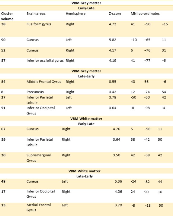
The parametric maps (color scale indicates Z scores, which correlate with the significance of the change) obtained from statistical analyses are superimposed on three orthogonal MNI-spaced T1-weighted average images. Relative to the late blind subjects, the EB subjects showed decreased WM volume in bilateral optic radiation and reduced GM volumes in bilateral primary sensori-moter areas(Figure2). In LB1 group secondary visual cortical area and primary auditory area but in case of LB2 primary visual cortical area along with Broca's and Wernicke's area in primary auditory cortex have higher grey matter concentration (Figure 3). In both early blind group bilateral striate, extrastriate areas, Wernicke's area along with left middle occipital and middle frontal gyrus and an increase in White Matter volume was found in superior frontal gyrus, left middle temporal gyrus was observed.Discussion
The observation that the EB subjects show atrophy of the optic radiation. Our results, however, are consistent with the findings in blind subjects, who reportedly show preserved GM volume and reduced WM volume in the superior temporal gyrus in the early auditory region. We observed WM volumes and decreased GM volumes in the primary senserimotor areas of our early blind subjects. This result is surprising given that learning- and/or overuse-induced plasticity in the brain is often associated with increases, rather than decreases, of GM volumes, in the brain regions involved [2,3]. In the early blind individuals, the white matter plasticity during the critical developmental period may compensate for the impairment resulting from the de-afferentation of the visual information that may explain for the early blind subjects show fewer regions with white matter impairments relative to the late blind.Acknowledgements
DST for Funding the studyReferences
1. Vos P et al., Brain. 2014;137:1224-40 2. Hou F et al., Biomed Res Int. 2017;2017:6756927 3. Modi S et al., Eur J Radiol. 2012;81:2811-9Figures
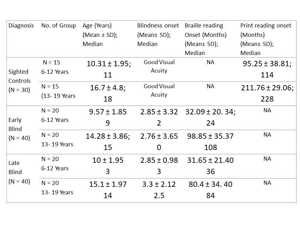
Sample
Size Detail (p1 = 0.45; p2 = 0.34)

Voxel-based morphometry analysis of group effects on regional brain gray matter, red regions denoting areas with lower grey matter volume in early blind (EB1, EB2) and late blind (LB1 and LB2) group when compared to sighted controls (SC1 and SC2).
Regions with significant change in gray matter volume in early blind and late blind subjects