3770
BMI correlates with tissue stiffness in deep gray matter regions controlling eating behavior1Berlin Center for Advanced Neuroimaging, Charité - Universitätsmedizin Berlin, Berlin, Germany, 2Bernstein Center for Computational Neuroscience, Berlin, Germany, 3Cluster of Excellence NeuroCure Clinical Research Center, Charité - Universitätsmedizin Berlin, Berlin, Germany, 4Institute of Medical Informatics, Charité - Universitätsmedizin Berlin, Berlin, Germany, 5Institute of Radiology, Charité - Universitätsmedizin Berlin, Berlin, Germany
Synopsis
Cerebral MR elastography was applied to a group of healthy male subjects in the normal weight range up to overweight to explore a potential relationship between the in vivo mechanical properties of brain tissue and body-mass index (BMI). We observed a highly significant negative correlation between tissue stiffness and BMI in the globus pallidus and putamen – two regions identified in the literature as being linked to eating behaviour – while the stiffness of other brain regions did not correlate with BMI. This is the first report on in vivo mechanical properties of brain tissue related to BMI.
Introduction
MR elastography (MRE) allows the non-invasive measurement of in vivo brain viscoelastic properties [1]. The interaction between tissue microstructure, microvascular blood flow and mechanical state of in vivo tissues offers a variety of quantitative imaging biomarkers for the assessment and better understanding of physiological and pathological processes in the brain. Cerebral MRE has recently been demonstrated to be sensitive to the variation of stiffness among deep gray matter regions in healthy human subjects with sensitivity to blood perfusion and vascular density [2]. Furthermore, the mechanical state of in vivo tissues has recently been shown to be sensitive to pathological processes such as neuroinflammation [5] and neurodegeneration [7].
In this study, we applied cerebral MRE to investigate whether tissue mechanical properties of the brain are also related to markers of metabolic disorders, i.e. the body-mass index (BMI). The BMI is strongly related to structural and functional neural mechanisms of reward and motivation processing localized in deep gray matter (DGM) regions. Specifically, it has been shown that DGM regions are linked to dopamine D2 receptor availability assessed with PET [3], as well as to food-cue responsivity in the striatum assessed with task-based fMRI [4].
Our hypothesis is that variations of tissue structure in striatal regions reflected by MRE parameters correlate with the variation of BMI among healthy subjects.
Methods
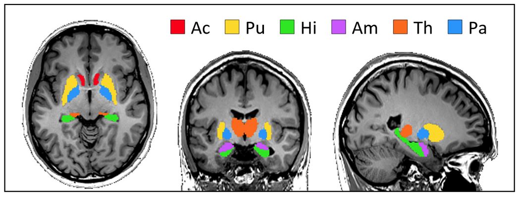
22 healthy male subjects (mean age 35, range 18 to 57 years) with normal weight and overweight (mean BMI 25, range 19 to 28 kg/m2) were included in this study. Cerebral MRE was performed in a 3-T MRI scanner (Siemens, Trio) using a custom-designed head cradle actuator for inducing mechanical vibrations at 30, 40, and 50 Hz [2]. Further MRE imaging parameters: 40 transversal slices of 2 mm isotropic resolution, TE=82ms, TR=8.49s, 8 acquisitions over a wave cycle. T1w-anatomical imaging (MPRAGE) with full brain coverage was applied prior to MRE. MRE postprocessing was based on multifrequency direct inversion [2,5] and combined with correction of field distortions [6]. 3D elastography maps reflecting the magnitude of the complex shear modulus |G*| were then normalized to fit the dimensions of the tissue probability maps and the Neuromorphometrics atlas of brain regions in MNI152 space to allow for inter-subject statistics. Six subcortical regions of interest (ROI) shown in figure 1 were analyzed according to [2].Results
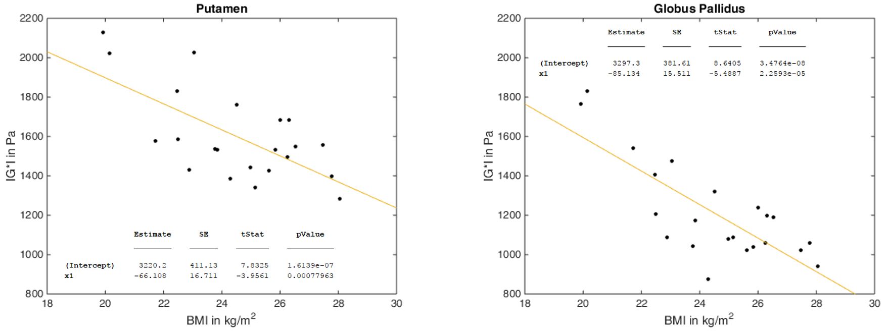
The analysis linking region-wise averages of tissue elasticity to BMI showed that stiffness in putamen (t=-4.0, p=8×10⁻⁴) and globus pallidus (t=-5.5, p=2×10⁻⁵) was significantly correlated to the BMI on a family-wise error corrected significance level (see figure 2). Contrarily, stiffness values of nucleus accumbens (t=0.4, p=0.7), hippocampus (t=-1.2, p=0.3), amygdala (t=-0.08, p=0.9) and thalamus (t=-2.0, p=0.06) were not significantly correlated to BMI.
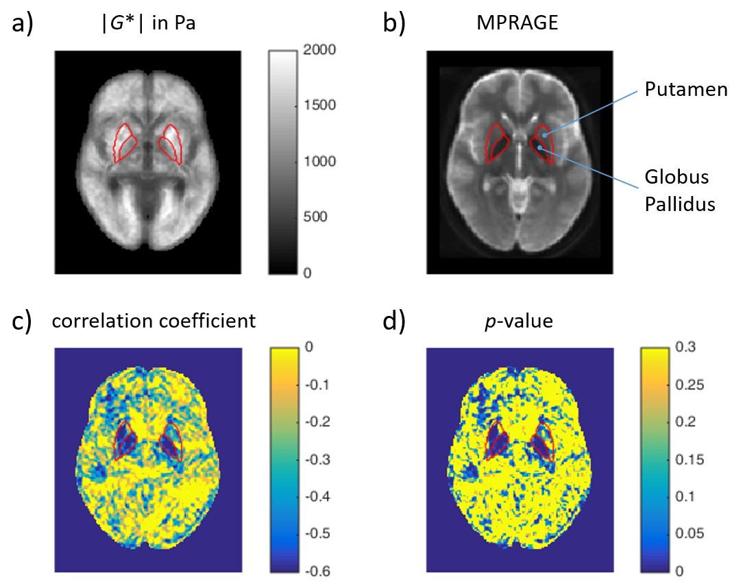
Additionally, figure 3 illustrates the spatial representation of the correlation between stiffness parameters and BMI und suggests the strong link between stiffness and BMI in globus pallidus and putamen observed by our region-wise analyses (see figure 2).
Discussion
A variety of behavioral parameters are known to influence BMI which can either be cause or consequence of tissue structural changes in specific brain regions. The putamen and globus pallidus are known as regions of high dopamine activity in close relationship to eating behavior. Our study shows for the first time that individual variations of tissue structure in that region can be measured by MRE and correlated with BMI values. This indicates that the relatively large variation of MRE-measured stiffness values within groups of healthy subjects as reported in the MRE literature might be related to physiological variations of brain matter [1]. The high sensitivity of MRE to such variations might be explored in the future for a better understanding of the relationship between tissue structure, brain function and individual behavior.Acknowledgements
No acknowledgement found.References
[1] Hiscox et al., Phys Med Biol 2016; 61:401-437
[2] Hetzer et al., JCBFM 2017; doi:10.1177/0271678X17691530
[3] Wang et al., Lancet 2001; 357:354-357
[4] Rothemund et al., NeuroImage 2007; 37:410-421
[5] Fehlner et al., JMRI 2016; 44:51-58
[6] Fehlner et al., JMRI 2017; 46:134-141
[7] Murphy et al., NeuroImage Clin 2015; 10:283-290
Figures