3736
Changes in brain connectivity of the first-episode idiopathic epilepsy patients before and after treatment: A comparative study of resting-state fMRIPengfei Qiao1, Guang-ming Niu1, Yang Gao2, and Lizhi Xie3
1Department of Radiology, Affiliated Hospital of Inner Mongolia Medical University, Hohhot, China, 2Department of Radiology, he Affiliated Hospital of Inner Mongolia Medical University, Hohhot, China, 3GE Healthcare, China, Beijing, China
Synopsis
This study investigates the fractional amplitude of low frequency fluctuation (fALFF) of the first-episode complex partial seizures epilepsy patients before and after treatment by using resting state fMRI (rfMRI). It is found that fALFF can be used to detect interictal epileptiform change before and after treatment.
Purpose
To investigate the changes of fractional amplitude of low frequency fluctuation (fALFF) in the first-episode complex partial seizures (CPS) epilepsy patients before and after treatment.Material and Methods
Fourteen right-handed CPS patients were enrolled in the comparison with fourteen matched controls in age, gender and education background. All subjects underwent MR imaging on a 3.0 T scanner (GE-Discovery 750, Milwaukee, US.). Functional MRI was performed using GRE-EPI sequences (FOV 24 cm × 24 cm, 64×64 matrix, flip angle 90°, TR/TE: 2000ms/ 30ms). The whole brain was covered with 38 oblique axial, 4mm slices thickness without gap. Resting-state scanning lasted for 512s, producing 256 brain volume data sets. The first 10 images were excluded due to T1 equilibrium effects. T1-weighted 3DBRAVO-sequence images (FOV 24 cm×24 cm, 256×256 matrix, whole brain coverage, flip angle 13°, TR/TE: 7.8ms/3.0ms) were achieved to assess the anatomical structure for the co-registration of fMRI data with standard space coordinates. After preprocessing (SPM8), the frame-wise displacement (FD) was controlled to be less than 0.5 mm to reduce the effect of motion. Volumes with FD > 0.5 mm were removed and subjects with at least 148 volumes were included in this study. fALFF were analyzed by using REST to compare the resting state function in the whole brain between two groups.Results
Before treatment, the CPS group showed significantly higher fALFF in the bilateral thalami, posterior cingulate, left angular gyrus and inferior parietal lobule, and significantly reduced fALFF in the bilateral medial prefrontal lobes and anterior cingulate compared with the control group. After treatment, abnormally activated areas were reduced or disappeared in the thalamus and angular gyrus, and increased in the left inferior parietal lobule. In addition, newly activated brain areas were detected in the bilateral frontal lobes, precuneus, and left cuneus (Figure1, 2; Tables 1, 2 and 3).Conclusion
The alterations of amplitude are observed in epileptogenesis and the change of fALFF is detected in the interictal epileptiform before and after treatment. Accordingly, fALFF might be used as an additional non-invasive tool for the detection of epileptogenic foci. The rsMRI provides new insights into the outcome evaluation, the effect of anti-epileptic drugs and the pathophysiological mechanism of epilepsy.Acknowledgements
No acknowledgement found.References
[1] Zou QH, Zhu CZ, Yang Y, et al: An improved approach to detection of amplitude of low-frequency fluctuation ( ALFF) for resting-state fMRI: fractional ALFF. J Neurosci Methods, 2008; 172: 137-141.
[2] Qiao PF, Niu GM: The application of ALFF to the idiopathic complex partial seizures epilepsy of teenagers. Neurology, psychiatry and brain research, 2015; 21: 27-32.
Figures
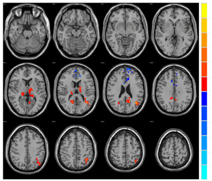
Figure.1 Two sample t-test on fALFF
value, maps showing statistically significant different regions in the group
comparisons of the CPS before treatment group compared with the control group
(increased fALFF labeled in warm color, decreased fALFF labeled in cold color).
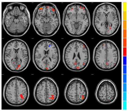
Figure.2 Group comparison between
the CPS with the control after treatment, abnormally activated areas were reduced or
disappeared in the thalamus and angular gyrus, and increased in the left
inferior parietal lobule. In addition, newly activated brain areas were
detected in the bilateral frontal lobes, presumes, and left cuneus.
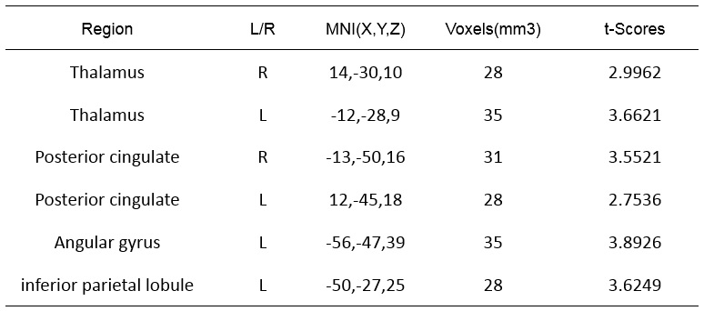
Table.1
Regions showing increased fALFF in the group comparisons between the before
treatment group and healthy group.
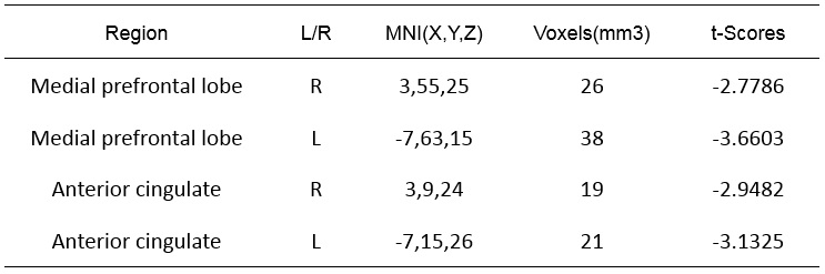
Table.2 Regions
showing decreased fALFF in the group comparisons between the before treatment
group and healthy group.

Table.3
Regions showing increased fALFF in the group comparisons between the after-treatment
group and healthy group.