3726
Gray matter cortical thickness changes in Hypothyroid patients: A study using high-resolution structural imaging1NMR Research Center, Institute of Nuclear Medicine and Allied Sciences, New Delhi, India, 2Thyroid Research Center, Institute of Nuclear Medicine and Allied Sciences, New Delhi, India
Synopsis
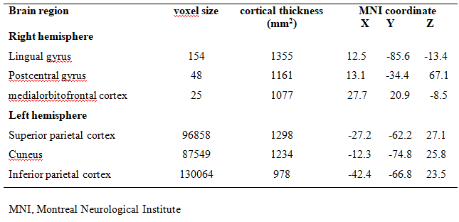
The aim of our study was to assess changes in cortical thickness, cortical area and cortical volume of gray matter (GM) for hypothyroid patients. We had acquired high-resolution 3D T1 weighted structural data for both control (24) and hypothyroid subjects (22). Reduced gray matter cortical thickness was observed in lateraloccipital cortex, postcentral gyrus, medialorbitofrontal cortex, lingual gyrus, superior and inferior parietal cortex in hypothyroid patients as compare to controls. These findings of reduced gray matter (CTh) suggest abated activities of motor, attention, working memory and executive cognitive function in hypothyroid patients.
Introduction
Hypothyroidism is defined as elevated levels of thyroid-stimulating hormone (TSH) and low levels of tri-iodothyronine (T3) and thyroxine (T4). Thyroid hormones (THs) play an important role in the normal functioning of the human brain 1. The lack of THs during human brain development leads to irreversible mental retardation and neurological deficits 1. In adulthood, THs are involved in the maintenance of brain structure and functionalities 2,3. The anomaly in levels of THs in adults reflects impaired cognitive functions and neuropsychiatric problems. In order to measure changes in cortical thickness, cortical area and cortical volume of gray matter (GM) for hypothyroid patients we have used high-resolution structural imaging data.Material and method
24 control subjects (mean age ± SD = 31.75 ± 8.60) and 22 hypothyroid patients (mean age ± SD = 31.94 ± 10.49) participated in the study. The clinical reports of hypothyroid patients show decreased FT4 and FT3 levels and increased TSH (>10 μIU/ml). None of the subjects had any history of neurological or psychiatric disorders. The study was approved by the institutional ethics committee. High-resolution 3D T1 weighted neuro-imaging data was obtained using 3T whole-body MR system (Magnetom; Siemens, Skyra, Germany) with a circularly polarized 20 channel matrix head and neck coil and a 45mT/m actively shielded gradient system using three-dimensional gradient echo sequence (Magnetization-Prepared Rapid Acquisition Gradient Echo, 160 sagittal slices, slice thickness = 0.9 mm, field of view = 240 mm, TR = 1900 ms, TE = 2.49 ms). We accomplished data analysis and data visualization using automated streamlines of FreeSurfer 5.6.0 software library (http://surfer.nmr.mgh.harvard.edu). The image analysis steps included motion correction, Talairach transformation, removal of non-brain tissues, and segmentation into GM/WM followed by deformation, cortical inflation, registration to a spherical atlas, and parcellation of the cerebral cortex into gyral and sulcal areas. Cortical thickness (CTh) was calculated as the closest distance from the pial to WM boundary at each vertex and surface area computed at the interface between gray matter (GM) and white matter (WM) 4. CTh and surface area maps were spatially smoothed with a Gaussian kernel with a full width at half maximum (10 mm) 4. We used a general linear model to compare maps of CTh and surface area between hypothyroid patients and healthy controls, the results are reported at p < 0.05 corrected for multiple comparisons using Monte-Carlo simulations.Result and Discussion
Our result showed significant decrease in cortical thickness lateral-occipital cortex, postcentral gyrus, medial orbitofrontal cortex, lingual gyrus in right hemisphere and cuneus, superior and inferior parietal cortex of the left hemisphere in hypothyroid patients as compared to controls (given Table 1 and Figure 1). The reduced CTh in hypothyroid patients suggests cognitive function decline associated brain region. The reduced CTh in postcentral gyrus suggests motor function deficit in hypothyroid 5. Reduced CTh in medial orbitofrontal cortex suggests impairment in working memory, problem-solving and decision-making ability in hypothyroid patients as compared to controls 6. Whereas reduced CTh in lingual gyrus, superior and inferior parietal cortex may be suggesting a decline in executive function, emotion, attention and concentration cognitive function in hypothyroid patients 6,7.Conclusion
Reduced gray matter cortical thickness was observed in lateral-occipital cortex, postcentral gyrus, medial orbitofrontal cortex, and lingual gyrus, superior and inferior parietal cortex. The reduced gray matter CTh suggest a cognitive decline in hypothyroid patients.Acknowledgements
We acknowledge that this research was supported by 'Defense Research and Development Organization' (DRDO), Ministry of Defense, Government of India.References
- Anderson GW. Thyroid hormone and cerebellar development. The Cerebellum. 2008; 7(1):60-74.
- GE, Mullally S, Correia N, O'Mara SM, Gibney J. Hippocampal volume is decreased in adults with hypothyroidism. Thyroid. 2014; 24(3):433-40.
- Singh S, Modi S, Bagga D, Kaur P, Shankar LR, Khushu S. Voxel‐Based Morphometric Analysis in Hypothyroidism Using Diffeomorphic Anatomic Registration via an Exponentiated Lie Algebra Algorithm Approach. Journal of neuroendocrinology. 2013;25(3):229-34.
- Fischl B, Dale AM. Measuring the thickness of the human cerebral cortex from magnetic resonance images. Proceedings of the National Academy of Sciences. 2000;97(20):11050-5.
- Khushu S, Kumaran SS, Sekhri T, Tripathi RP, Jain PC, Jain V. Cortical activation during finger tapping in thyroid dysfunction: a functional magnetic resonance imaging study. Journal of biosciences. 2006;31(5):543-50.
- He XS, Ma N, Pan ZL, Wang ZX, Li N, Zhang XC, Zhou JN, Zhu DF, Zhang DR. Functional magnetic resource imaging assessment of altered brain function in hypothyroidism during working memory processing. European Journal of Endocrinology. 2011;164(6):951-9.
- Blasi V, Longaretti R, Giovanettoni C, Baldoli C, Pontesilli S, Vigone C, Saccuman C, Nigro F, Chiumello G, Scotti G, Weber G. Decreased parietal cortex activity during mental rotation in children with congenital hypothyroidism. Neuroendocrinology. 2009;89(1):56-65
Figures
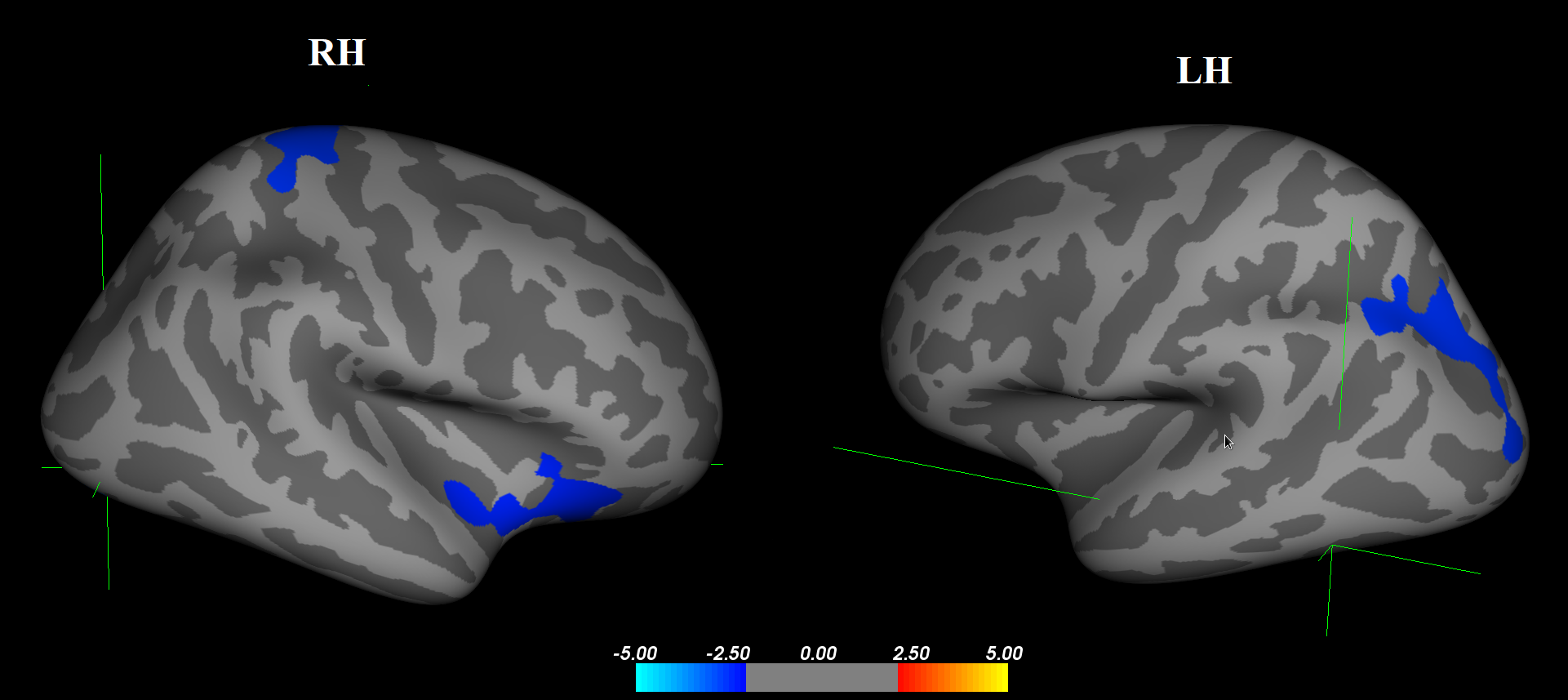
Cortical regions showing decrease in gray matter cortical thickness in hypothyroid patients as compared to healthy controls.
RH (Right hemisphere); LH (Left hemisphere)