3721
Brain microstructural abnormalities in cirrhotic patients without overt hepatic encephalopathy: A voxel-based diffusional kurtosis imaging study1Fujian Medical University Union Hospital, Fuzhou, China
Synopsis
Brain microstructural change in cirrhotic patients without overt hepatic encephalopathy: A DKI study
Introduction
Brain morphological abnormalities have been well identified in cirrhotic patients, whose condition concomitantly involves gray matter (GM) and white matter (WM) [1-4]. Diffusion tensor imaging (DTI) is the most frequently used MR diffusion-weighted technique for evaluating cirrhosis-related changes in the cerebral microstructure [3, 5-7]. Notably, all of these previous studies were based on the conventional DTI technique, which assumes the water diffusion to be a Gaussian distribution. As a natural extension of diffusion tensor imaging, the diffusional kurtosis imaging (DKI) method employs multiple b-values to quantify non-Gaussian diffusion by estimating the excess kurtosis of the displacement distribution, in which both standard DTI parameters and kurtosis parameters—including mean kurtosis (MK), axial kurtosis (K∥), and radial kurtosis (K⊥)—could be obtained. Thus, by taking into account the fundamentally non-Gaussian property of water diffusion in biological tissue, DKI can function as a complementary technique to the conventional DTI to more fully depict the characterization of brain tissue microstructure [8-11]. The kurtosis metrics are believed to reflect the heterogeneity of the intra-voxel diffusion environment and are thus indicators of microstructural complexity. Moreover, DKI permits the characterization of microstructural integrity in both GM and WM. Thus, DKI provides an opportunity for not only WM region exploration, as in previous DTI studies, but for whole-brain exploration of microstructural abnormalities in cirrhosis. DKI is thought to exhibit improved sensitivity and specificity in detecting changes in neural tissues [12]. Thus, the goal of the present study was to employ DKI to investigate the changes in both GM and WM microstructure in cirrhotic patients using a voxel-based analysis. The relationship between brain microstructural abnormalities and cognitive performance was also examined.Methods
Eighteen cirrhotic patients without overt hepatic encephalopathy (HE) and 17 healthy controls underwent diffusion imaging. There were no significant differences between control and patients groups, in terms of age, sex , or education level. Cognition was measured using Psychometric Hepatic Encephalopathy Score (PHES). Whole-brain voxel-based analyses (VBA) were performed to investigate between-group differences in DKI-derived parameters, including MK, K∥, and K⊥. The global GM and WM DKI-metrics were computed for each cirrhotic patient. Spearman correlation analyses were performed to measure the relationships between the DKI variables and cognitive performance in cirrhotic patientsResults
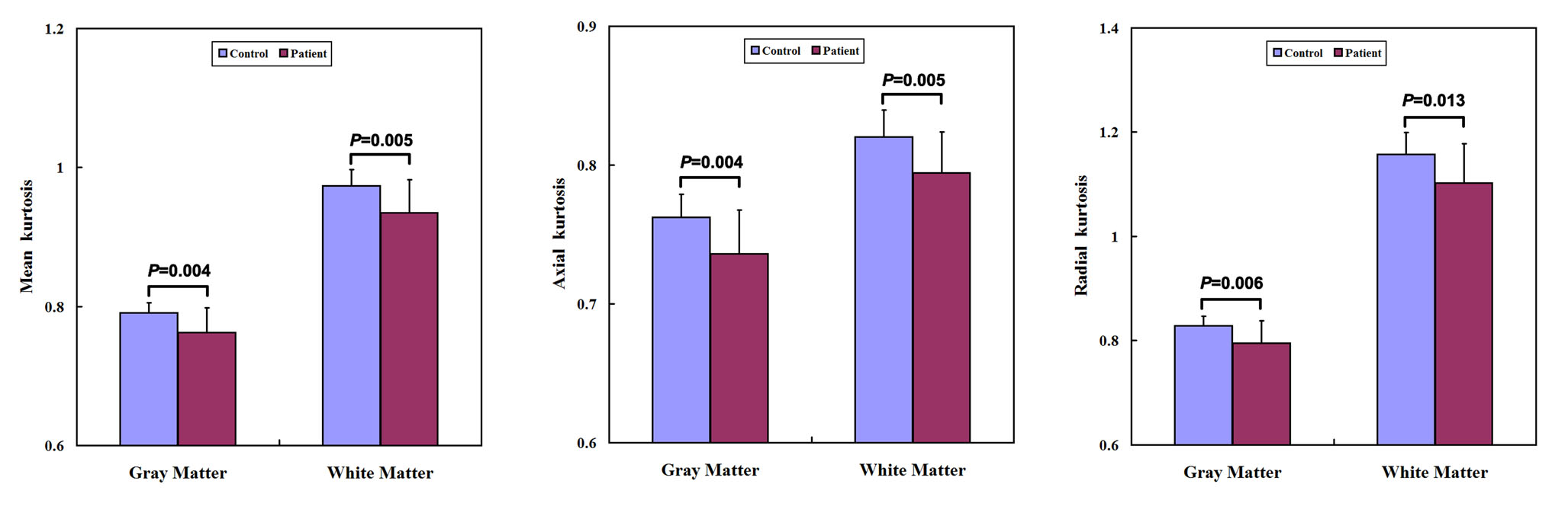
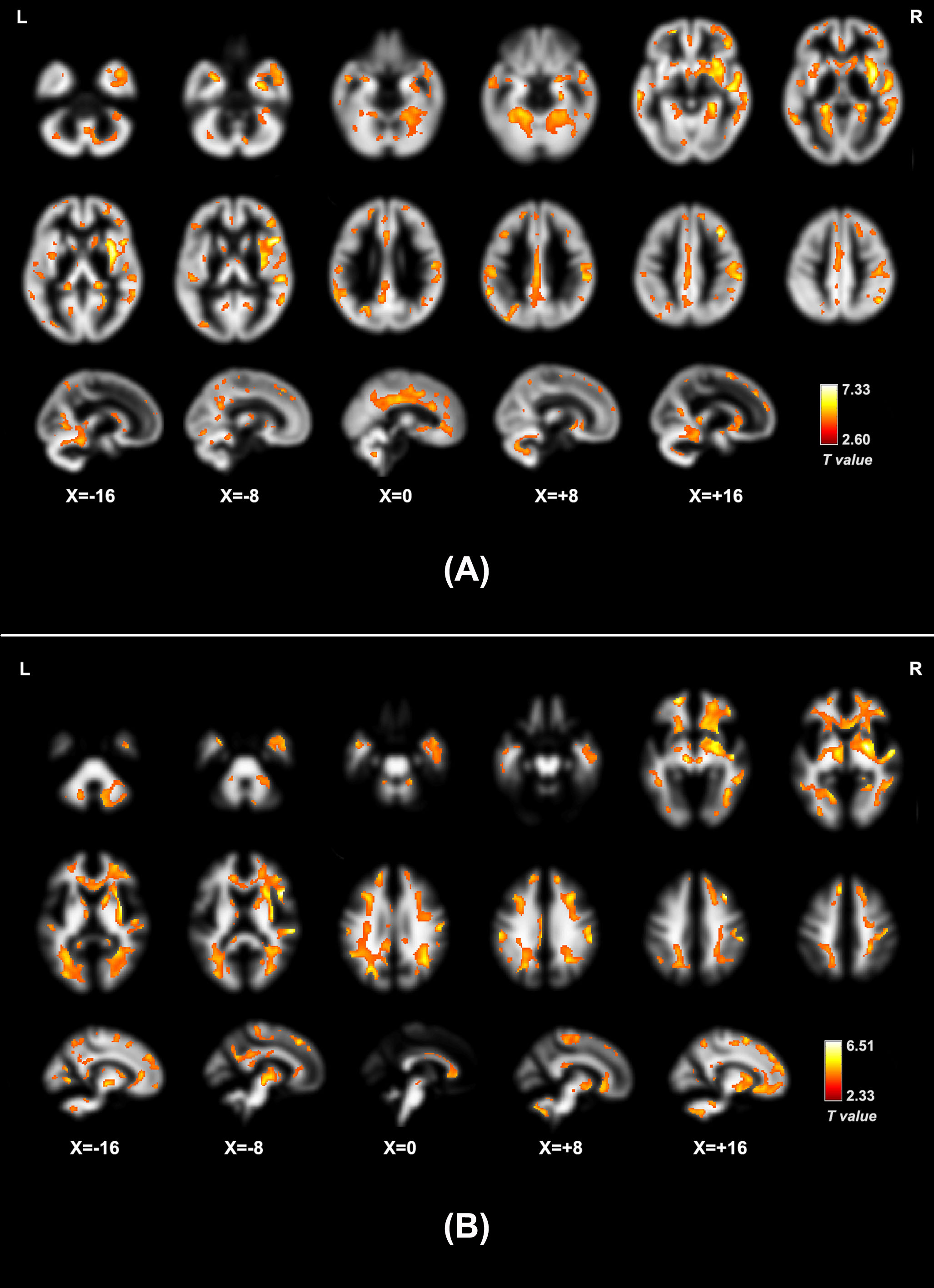
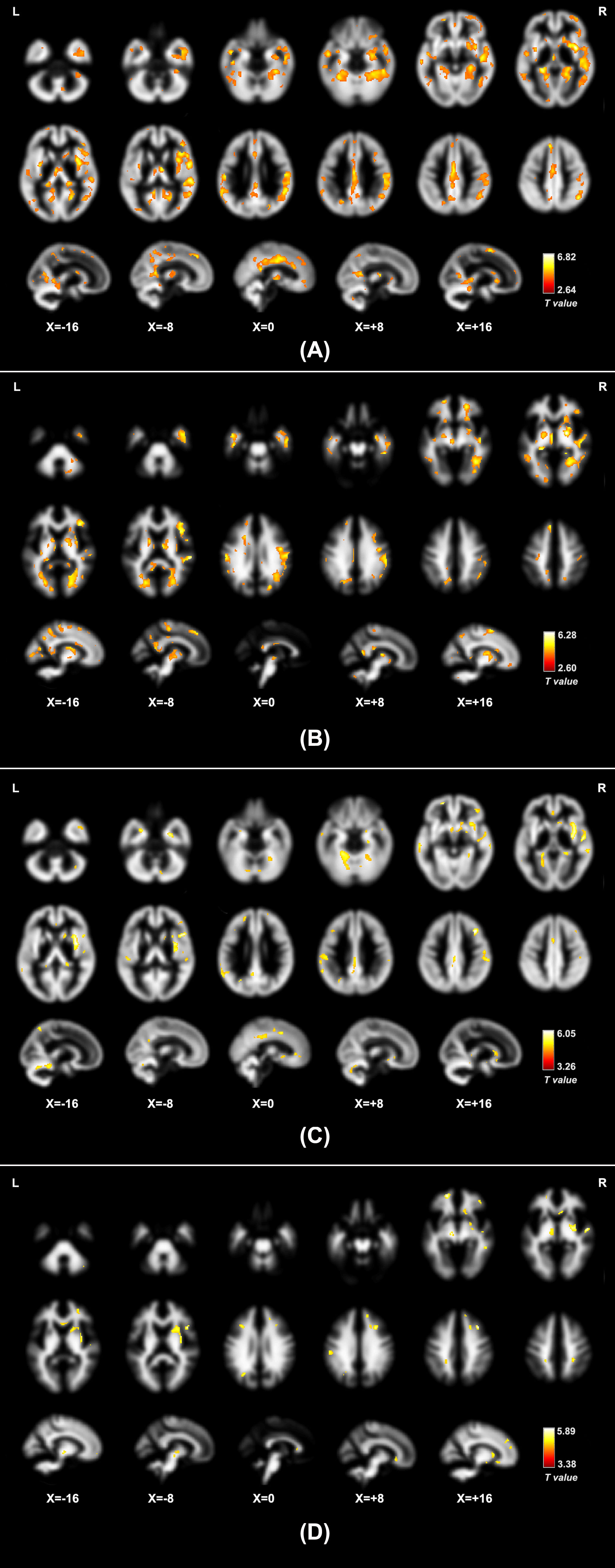
The cirrhotic patients had worse performance in all five PHES subtests and lower PHES score than the healthy controls (-3.3 ± 4.8 vs 0.6 ± 1.6, P = 0.004), indicating cognitive impairments. The cirrhotic patients had significantly lower global MK, K∥, and K⊥ in GM and WM (Figure 1). VBA showed that cirrhotic patients had decreased MK, K∥, and K⊥ in diffuse GM regions (particularly in cingulate cortex, precuneus, insular cortex, frontal areas, basal ganglia, hippocampus/ parahippocampal gyrus, supramarginal gyrus and angular gyrus, postcentral and precentral gyrus, and cerebellum) and WM regions (particularly in corpus callosum, internal capsule, frontal regions, parietal regions, occipital regions, and cerebellum) (Figures 2-3). The correlation analysis showed that the average GM MK (r = 0.474, P = 0.047), K∥(r = 0.470, P = 0.048), and K⊥ (r = 0.495, P = 0.039) were positively correlated with PHES score in cirrhotic patients. Also, the mean WM MK (r = 0.581, P = 0.011), K∥(r = 0.501, P = 0.034), and K⊥ (r = 0.615, P = 0.007) were significantly correlated with PHES score.Discussion
The DKI parameter, MK, is usually thought to reflect the microstructural complexity of brain tissues (GM microstructural complexity may be associated with dendrites/spines, synaptic pruning/refinement, and cell packing density, while WM microstructural complexity may be associated with myelination, denser axon and fiber bundle packing, refined fiber organization, and axonal membrane permeability [13]). Lower global MK parameter was found in both GM and WM in patients, indicating decreased microstructural complexity. The diffuse reduction of GM and WM microstructural complexity and its relationship with cognitive impairment in cirrhotic patients were revealed by our DKI study, and the results were in line with previous reports [1-3, 7,14-15]. In addition, the reduction of K∥ and K⊥ may indicate the impairment of axon function and myelination in WM related to cirrhosis, repetitively.Conclusion
The present study outlined whole-brain microstructural abnormalities in cirrhotic patients without overt HE, which were diffusely distributed in both GM and WM regions. Lower diffusional kurtosis parameters suggest decreased brain microstructural complexity in cirrhotic patients. The altered microstructural properties of both GM and WM were correlated with the cognitive performance of patients, suggesting the potential of these metrics to serve as biomarkers for early diagnosis of HE. DKI can provide supplementary information for characterizing brain structural abnormalities in cirrhosis.Acknowledgements
This work was supported by a grant from the National Natural Science Foundation of China (No. 81501450) and funding from the China Postdoctoral Science Foundation.References
1. Chen HJ, Wang Y, Zhu XQ, et al. White matter abnormalities correlate with neurocognitive performance in patients with HBV-related cirrhosis. J Neurol Sci. 2012;321:65-72.
2. Guevara M, Baccaro ME, Gomez-Anson B, et al. Cerebral magnetic resonance imaging reveals marked abnormalities of brain tissue density in patients with cirrhosis without overt hepatic encephalopathy. J Hepatol. 2011;55:564-573.
3. Montoliu C, Urios A, Forn C, et al. Reduced white matter microstructural integrity correlates with cognitive deficits in minimal hepatic encephalopathy. Gut. 2014;63:1028-1030.
4. Amodio P, Pellegrini A, Amista P, et al. Neuropsychological-neurophysiological alterations and brain atrophy in cirrhotic patients. Metab Brain Dis. 2003;18:63-78.
5. Chen HJ, Wang Y, Zhu XQ, et al. White matter abnormalities correlate with neurocognitive performance in patients with HBV-related cirrhosis. J Neurol Sci. 2012;321:65-72.
6. Qi R, Zhang LJ, Zhong J, et al. Grey and white matter abnormalities in minimal hepatic encephalopathy: a study combining voxel-based morphometry and tract-based spatial statistics. Eur Radiol. 2013; 23(12): 3370-3378.
7. Kumar R, Gupta RK, Elderkin-Thompson V, et al. Voxel-based diffusion tensor magnetic resonance imaging evaluation of low-grade hepatic encephalopathy. J Magn Reson Imaging. 2008;27:1061-1068.
8. Jensen JH, Helpern JA, Ramani A, et al. Diffusional kurtosis imaging: the quantification of non-gaussian water diffusion by means of magnetic resonance imaging. Magn Reson Med. 2005;53:1432-1440.
9. Jensen JH, Helpern JA. MRI quantification of non-Gaussian water diffusion by kurtosis analysis. NMR Biomed. 2010;23:698-710.
10. Lu H, Jensen JH, Ramani A, et al. Three-dimensional characterization of non-gaussian water diffusion in humans using diffusion kurtosis imaging. NMR Biomed. 2006;19:236-247.
11. Wu EX, Cheung MM. MR diffusion kurtosis imaging for neural tissue characterization. NMR Biomed. 2010;23:836-848.
12. Cheung MM, Hui ES, Chan KC, et al. Does diffusion kurtosis imaging lead to better neural tissue characterization? A rodent brain maturation study. Neuroimage. 2009;45:386-392.
13. Adisetiyo V, Tabesh A, Di Martino A, et al. Attention-deficit/hyperactivity disorder without comorbidity is associated with distinct atypical patterns of cerebral microstructural development. Hum Brain Mapp. 2014;35:2148-2162.
14. Zhang LJ, Qi R, Zhong J, et al. The effect of hepatic encephalopathy, hepatic failure, and portosystemic shunt on brain volume of cirrhotic patients: a voxel-based morphometry study. PLoS One. 2012;7:e42824.
15. Qi R, Zhang LJ, Zhong J, et al. Grey and white matter abnormalities in minimal hepatic encephalopathy: a study combining voxel-based morphometry and tract-based spatial statistics. Eur Radiol. 2013;23:3370-3378.
Figures