3707
Imaging Regional Distribution of Metabolic Activity During Progression of Acute Lung Injury using Hyperpolarized Carbon-13 MRI1Radiology, University of Pennsylvania, Philadelphia, PA, United States, 2Anesthesiology and Critical Care, University of Pennsylvania, Philadelphia, PA, United States, 3Physiology, University of Pennsylvania, Philadelphia, PA, United States
Synopsis
We used hyperpolarized [1-13C] pyruvate MRI to assess regional alterations in lactate production during progression of acute lung injury. While the average lactate-to-pyruvate ratio and its standard deviation increased globally in lungs ventilated without recruitment, the average ratio increased more significantly in the posterior regions than the average ratio in the anterior region. The average lactate-to-pyruvate remained unchanged in lungs ventilated with recruitment maneuver. Our finding suggests that stretch-induced atelectasis and inflammation are the root of increased lactate-to-pyruvate ratio in the lungs ventilated without recruitment.
Introduction
Regional distribution of metabolism in injured lungs is not well characterized (1). While a number of studies using 18F-FDG-PET have shown high FDG uptake in dependent regions co-localized with atelectasis and inflammatory activation (2), others reported accelerated glucose metabolism in non-dependent lung regions exposed to high inspiratory stretch rather than atelectasis (1,3). In this study, we used hyperpolarized [1-13C] pyruvate MRI in an acid instillation lung injury model followed by low-tidal volume ventilation with and without recruitment using positive-end expiratory pressure (PEEP). Our study showed that anaerobic metabolism, measured via lactate-to-pyruvate ratio, increases globally in the injured lungs; however, it increases significantly more in dependent lung regions co-localized with alveolar collapse and radiological infiltrates.Materials and Methods
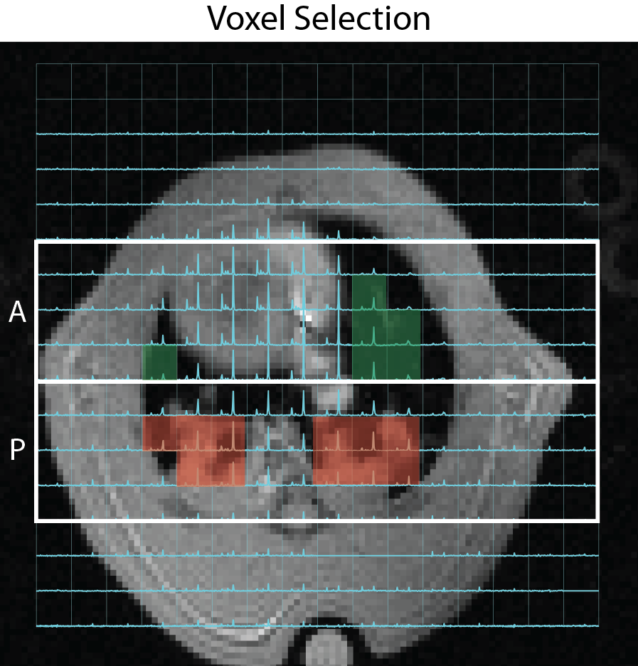
Fourteen Sprague-Dawley rats (308±10g) were ventilated (VT=8ml/kg, FiO2=1.0, PEEP=5cmH2O, frequency=52min-1). All rats were imaged in supine position using a dual tuned quadrature 1H/13C volume coil (m2m) in a 4.7T horizontal-bore small animal MRI system (Varian Inc.). Baseline axial and coronal proton scans and axial hyperpolarized [1-13C] pyruvate MRI was performed as previously described (4). All rats received intratracheal instillation of 0.5ml/kg hydrochloric acid (HCl, pH 1.25), after which ventilation was continued with the previous parameters. A second hyperpolarized [1-13C] pyruvate injection was performed 1 hour after acid instillation. Positive end expiratory pressure (PEEP) was then reduced to 0 cmH2O in seven injured rats (ZEEP group) to promote injury progression and alveolar collapse (atelectasis). PEEP was maintained at 5 cmH2O in the remaining rats (PEEP group) throughout the experiment. Two additional [1-13C] pyruvate injections were performed in both groups at 2.5 and 4 hours post-injury. Peak Inspiratory Pressure (PIP), pulmonary compliance (Cdyn) and oxygen saturation (SpO2) were monitored throughout the experiment to assess injury progression and ensure sufficient oxygenation (SpO2 > 90%). Regional analysis of the lactate-to-pyruvate ratio was performed using custom-made routines in MATLAB2015b by manually selecting hyperpolarized carbon-13 spectroscopic voxels covering the anterior and posterior regions of the lungs, as shown in Figure 1. Lactate-to-pyruvate ratio in each region was quantified from the average signal in these voxels. A histogram of the lactate-to-pyruvate ratio was generated from the ratio of individual voxels in each region.Results and Discussion
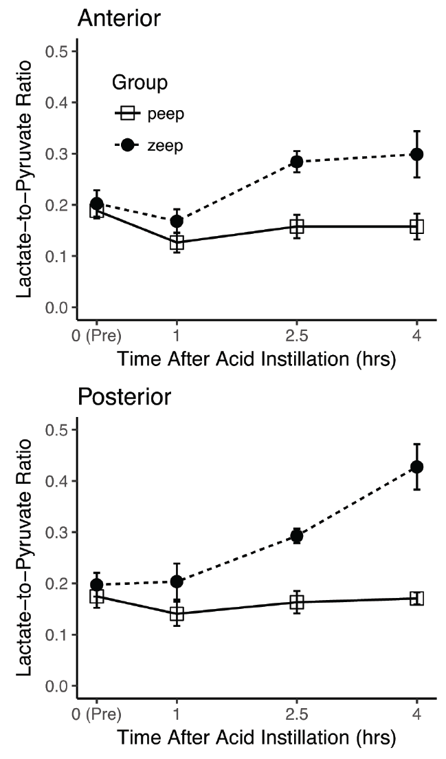
Figure 2 shows the trend of the average lactate-to-pyruvate ratio in anterior and posterior regions of both PEEP and ZEEP lungs. The ratio was similar in both regions of the lungs before and 1-hour after acid instillation, and it remained constant over time in the PEEP lungs. In the ZEEP lungs, however, the lactate-to-pyruvate ratio increased similarly in both anterior and posterior regions 2.5 hours after acid instillation. The ratio in the posterior region continued to increase 4 hours after acid instillation, while it remained unchanged and significantly lower in the anterior region (p=0.003).
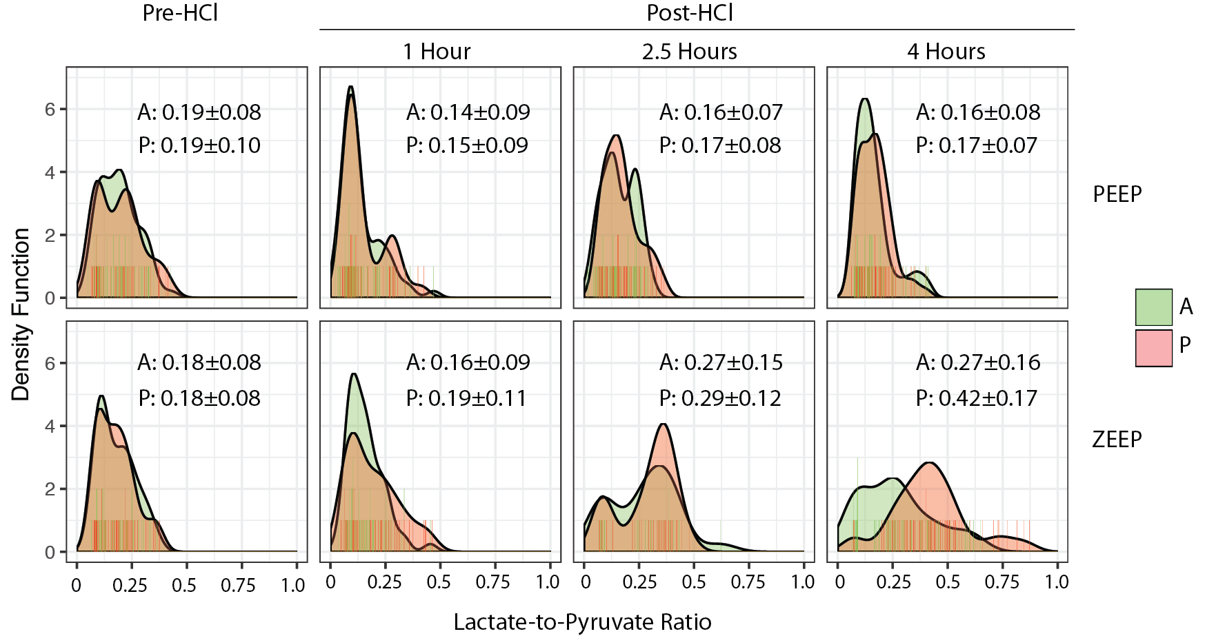
Figure 3 shows the smoothed histogram of the lactate-to-pyruvate ratio in the anterior and posterior regions of ZEEP and PEEP lungs. The standard deviation of the lactate-to-pyruvate ratio increased in the ZEEP lungs at both 2.5 and 4 hours after acid instillation, indicating increasingly heterogeneous metabolic activity in the lungs after injury progression. Interestingly, the ratio in both regions had a bimodal distribution 2.5 hours after acid instillation, suggesting presence of areas with normal metabolic activity in both regions. 4 hours after acid instillation, the distribution in the anterior region remained relatively unchanged. This distribution became unimodal in the posterior region, however, indicating increased anaerobic metabolism throughout the posterior lung due to injury progression.
While the lactate-to-pyruvate ratio increased in both regions, it increased
significantly more in dependent regions co-localized with areas displaying
increased proton density and more prominent radiological infiltrates. This
finding suggests that increased anaerobic metabolism in the absence of PEEP may
be related to dependent atelectasis. In fact, lactate-to-pyruvate ratio and its
histogram remained unchanged in both anterior and posterior lung regions in the
PEEP group, and was significantly lower than in ZEEP lungs at 2.5 and 4 hours
after acid instillation (p<0.001).
Conclusion
In this study, we demonstrated the utility of hyperpolarized [1-13C] pyruvate for assessing regional anaerobic lung metabolism in acute lung injury. We showed that the increase in lactate-to-pyruvate ratio is significantly higher in posterior regions where radiological infiltrates are more prominent. Such a regional distribution of the lactate and lactate-to-pyruvate signal in the absence of PEEP suggests abnormal tissue metabolism in lung regions with dependent atelectasis after lung injury in the absence of proper lung recruitment.Acknowledgements
No acknowledgement found.References
1. Bellani G, Guerra L, Musch G, Zanella A, Patroniti N, Mauri T, Messa C, Pesenti A. Lung Regional Metabolic Activity and Gas Volume Changes Induced by Tidal Ventilation in Patients with Acute Lung Injury. Am J Respir Crit Care Med 2011;183:1193–1199. doi: 10.1164/rccm.201008-1318OC.
2. Wellman TJ, Winkler T, Costa ELV, Musch G, Harris RS, Zheng H, Venegas JG, Vidal Melo MF. Effect of Local Tidal Lung Strain on Inflammation in Normal and Lipopolysaccharide-Exposed Sheep*. Critical Care Medicine 2014;42:e491–e500. doi: 10.1097/CCM.0000000000000346.
3. Tsuchida S, Engelberts D, Peltekova V, Hopkins N, Frndova H, Babyn P, McKerlie C, Post M, McLoughlin P, Kavanagh BP. Atelectasis Causes Alveolar Injury in Nonatelectatic Lung Regions. Am J Respir Crit Care Med 2006;174:279–289. doi: 10.1164/rccm.200506-1006OC.
4. Pourfathi M, Xin Y, Kadlecek SJ, et al. In vivo imaging of the progression of acute lung injury using hyperpolarized [1-(13) C] pyruvate. Magn. Reson. Med. 2017;307:2526. doi: 10.1002/mrm.26604.
Figures