3700
Hyperpolarized carbon-13 MRS of liver in a high-fat/high sugar diet guinea pig model1Medical Biophysics, Western University, London, ON, Canada, 2Robarts Research Institute, London, ON, Canada, 3Physiology and Pharmacology, Western University, London, ON, Canada, 4Obstetrics and Gynaecology, Schulich Medicine & Dentistry, London, ON, Canada, 5Maternal, Fetal & Newborn Health, Children's Health Research Institute, London, ON, Canada
Synopsis
Effects of a life long high-fat/high sugar diet (Western diet: WD) upon pyruvate liver metabolism were observed in a group of young adult male guinea pigs (N=26). Proton density fat-fraction images were reconstructed using IDEAL water fat separation. Metabolism data were obtained using dynamic spectroscopy of hyperpolarized carbon-13 enriched pyruvate. Guinea pigs fed a life long WD displayed a significantly higher hepatic fat fraction and a delayed time to peak for the conversion of pyruvate to lactate. These results indicate that life long consumption of a WD in growing animals is associated with markers of dysfunctional hepatic metabolic function.
Introduction
Hepatic metabolic dysfunction has been linked to the development of metabolic disease, including non-alcoholic fatty liver disease (NAFLD) and type II diabetes. Acute high fat feeding studies have demonstrated alterations in pyruvate metabolism.1 The aim of this study was to investigate how a life long Western (high-fat/high sugar) diet (WD) may impact hepatic metabolic function, specifically pyruvate metabolism. Techniques involving hyperpolarized magnetic resonance spectroscopy (MRS) allowed for the real-time quantification of central metabolic biomolecules such as pyruvate in vivo. Hyperpolarized carbon-13 (13C) MRS along with IDEAL fat-water imaging were used to acquire quantitative data related to liver function in young growing male guinea pigs fed a lifelong Western diet.Methods
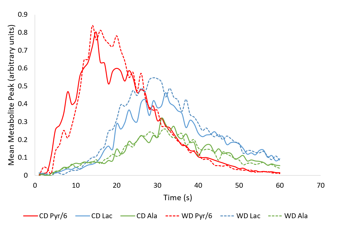
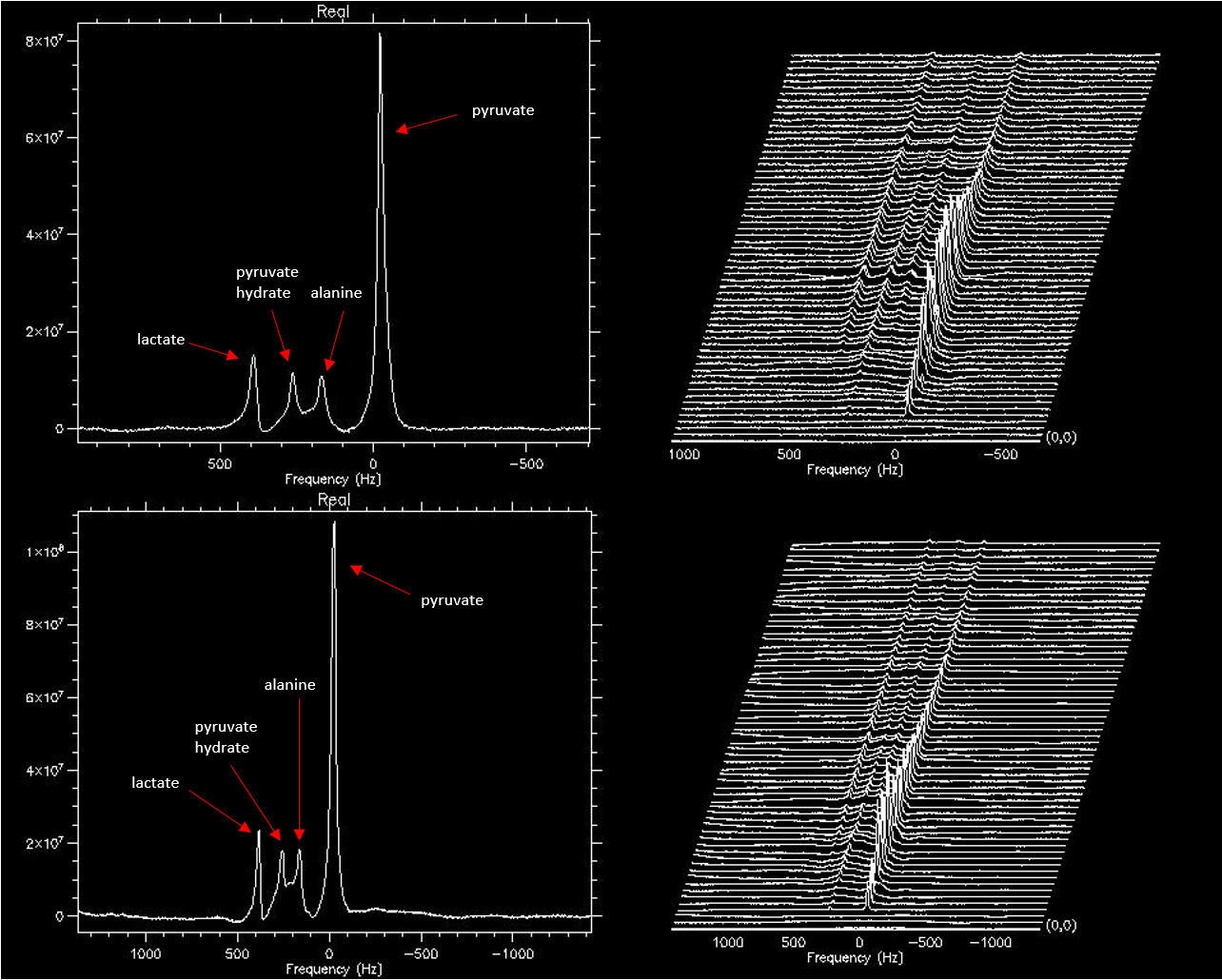
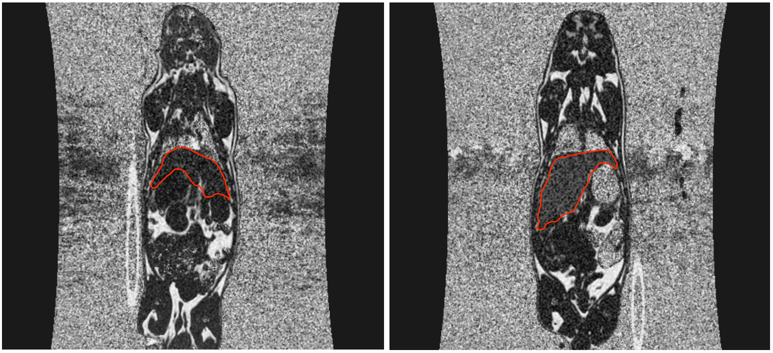
Normal birth weight male guinea pig pups (matched for litter) born in our facility were either weaned onto a Western diet (WD; 33% calories from carbohydrates, 46% fat, 21% protein, n=13) or a control diet (CD; 60% carbohydrates, 18% fat, 22% protein, n=13).2 After 144 +/- 4 days (young adulthood ~ 18-22 human years), animals were imaged using a 3T GE MRI under anaesthetic (1.5-2.5% isoflurane with 2L/min O2).2 T1 and T2 weighted images were acquired for anatomical reference and used to select slabs through the liver for 13C PRESS MRS. IDEAL images were acquired and reconstructed to create proton density fat-fraction (PDFF) images of the anatomy. Mean PDFF of ROIs drawn in the liver and hind leg muscle were calculated. Following bolus injection of hyperpolarized 13C labeled pyruvate, 13C spectra were acquired over 90 seconds with a 1 second time resolution. Time to peak (TTP) was measured as the time from the pyruvate peak to the metabolite peak. All TTPs were relative to the pyruvate peak to mitigate differences in injection times between animals.Results
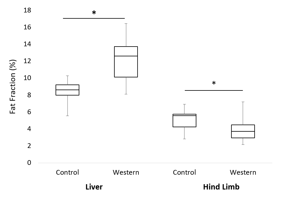
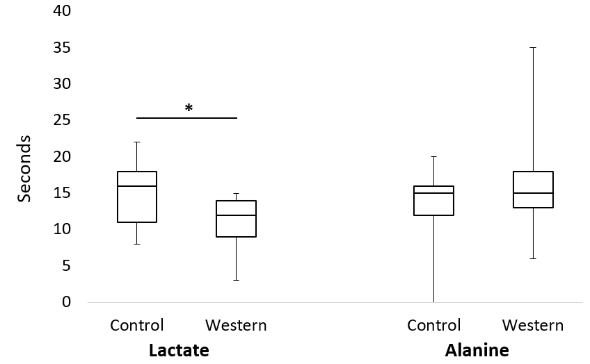
Measurement of fat fraction between groups showed a significant (p<0.05) increase in fat of the liver and decrease in hind leg fat for the Western diet group animals (Fig. 1). Hyperpolarized 13C spectroscopy MRI results showed a significant (p=0.01) decrease in TTP of lactate for the Western diet group (Fig. 4A). No significant differences were observed for the alanine TTP between diet groups (Fig. 4B). All statistical significance was calculated using a unpaired t-tests.Discussion
The increase of fat fraction in liver with a decline in muscle fat content is evidence that components of the WD promote differential fat deposition displayed as an increased hepatic fat deposition relative to muscle compared to the CD fed animals. This altered fat deposition pattern is associated with an altered pyruvate metabolic pathway where the time to peak for lactate is decreased in WD fed animals. TTP has been used as a model-free approach to estimate the metabolic conversion rate and is inversely related to the concentration of lactate dehydrogenase, the enzyme responsible for the metabolic conversion of pyruvate to lactate.3 It is likely that the Western diet animals were producing a greater amount of lactate, which is associated with anaerobic metabolic conditions.1 In theory, pyruvate should be entirely oxidized in the mitochondria during the TCA cycle to produce ATP.4 An increase in lactate production could indicate that a high-fat/high sugar diet may promote glycolytic metabolism. Previous literature4 has described altered liver metabolism as a precursor to metabolic diseases. Our data implies that lifelong exposure, from weaning into young adulthood, to high-fat/high sugar diets common to the Western world may be negatively impacting liver function early in life, which in turn predisposes a population to a higher incidence of metabolic disease with age.Conclusion
Hyperpolarized 13C MRI was shown to be a successful method for quantitative measurement of metabolic by-products in the liver of guinea pigs fed a lifelong WD. Determination of TTP for spectroscopic data allows for real-time information of metabolism within the liver. Combined with quantitative fat fraction imaging, measurement of these changes in metabolism of key nutrients, such as pyruvate, may provide early biomarkers that could be useful in identifying patients at risk of developing later life metabolic disease such as type II diabetes and cardiovascular disease. An observed decrease in lactate TTP for the Western diet animals indicates that a high-fat/high sugar diet can negatively influence metabolism in the liver. Future studies may consider investigating the animals in intervention regimes or studying the intergenerational effects of high-fat diets on future offspring’s metabolic health.Acknowledgements
No acknowledgement found.References
1. Moon, Chung-Man, et al. Metabolic biomarkers for non-alcoholic fatty liver disease induced by high-fat diet: In vivo magnetic resonance spectroscopy of hyperpolarized [1-13 C] pyruvate. Biochemical and biophysical research communications 482.1 (2017): 112-119.
2. Friesen‐Waldner, Lanette J., et al. Hyperpolarized [1‐13C] pyruvate MRI for noninvasive examination of placental metabolism and nutrient transport: A feasibility study in pregnant guinea pigs. Journal of Magnetic Resonance Imaging 43.3 (2016): 750-755.
3. Daniels, Charlie J., et al. A comparison of quantitative methods for clinical imaging with hyperpolarized 13C‐pyruvate. NMR in biomedicine 29.4 (2016): 387-399.
4. Rui, Liangyou. Energy metabolism in the liver. Comprehensive physiology (2014).
Figures