3688
CSF transport of 18F and 18FDG via the brain-wide glymphatic pathway visualized by Integrative PET-MRI1Anesthesiology, Yale School of Medicine, New Haven, CT, United States, 2Biomedical Engineering, Stony Brook Medicine, Stony Brook, NY, United States, 3SAII, Stony Brook, NY, United States, 4Radiology, Stony Brook Medicine, Stony Brook, NY, United States, 5NIAAA, Bethesda, MD, United States, 6University of Copenhagen, Copenhagen, Denmark
Synopsis
The glymphatic pathway was recently re-discovered as a CSF transport system for brain waste removal. The peri-vascular space functions as the ”front end” for toxic waste clearance via the ‘glymphatic pathway. To facilitate future translation to the clinic we executed experiments to map glymphatic transport in the live rodent brain using dynamic integrated MRI-PET imaging in combination with CSF administration of a paramagnetic contrast agent and two different 18F-labeled radioisotopes.
Introduction
In most of the body, the lymphatic system facilitates clearance of metabolic waste products and excess fluid. The brain parenchyma, however, is different. Devoid of authentic lymphatic vessels, the fundamental process by which waste generated in the brain is effectively removed has remained unclear. Recently, a previously underappreciated but unique component of the brain vasculature, the peri-vascular space, was discovered to function as the ”front end” for a toxic waste clearance process now known as the ‘glymphatic pathway’1. At present, anatomy of the glymphatic pathway is complex and only partly understood. The outer perimeter is defined by glial endfeet with high expression of aquaporin 4 (AQP4) water channels that facilitate convectively-driven CSF movement into the interstitial fluid space. Continuous propulsion of CSF from the peri-vascular space into the interstitial fluid (ISF) compartment helps drive soluble metabolic waste products including amyloid beta (Aβ) and tau peptides into peri-venous conduits for downstream removal2. Ultimately, peri-venous exit pathways for brain waste drain into authentic lymphatic vessels recently discovered in the meninges3 as well as lymphatic vessels along cranial nerves that drain to cervical lymph nodes. To facilitate future translation to the clinic we executed experiments to map glymphatic transport in the live rodent brain using dynamic MRI-PET imaging in combination with CSF administration of a paramagnetic contrast agent and two different 18F-labeled radioisotopes.Methods
Sprague Dawley rats were anesthetized with phenobarbital and CSF catheters placed. Imaging was performed using a custom-built, MRI-compatible PET scanner for the rat brain in combination with a 9.4T microMRI4. The rats were imaged in the supine position and a mixture of Gd-DTPA and 18F tagged isotopes were infused into CSF. Dynamic MRI and PET images were acquired synchronously; including a 3D T1-weighted FLASH sequence (TR=15msec, TE=3.8ms, NA=1, FOV=3.0x3.0x3.2 cm, scanning time = 4.1 min, image resolution of 0.12x0.12x0.13 mm) and PET acquisition in singles listmode, spatial resolution of 1.2mm. Two different 18F tagged isotopes mixed with Gd-DTPA (20.8mM in saline) were tested: 1) 0.5mCi [18F]fluoride and 2) 0.5mCi 2-deoxy-2-(18F)fluoro-D-glucose (18FDG). MRI data were reconstructed as previously described3. PET data was reconstructed using an iterative, maximum likelihood by expectation maximization (MLEM) approach and included the final corrections for decay and live time. PET-MRI image fusion and extraction of time-activity curves was executed using PMOD.Results
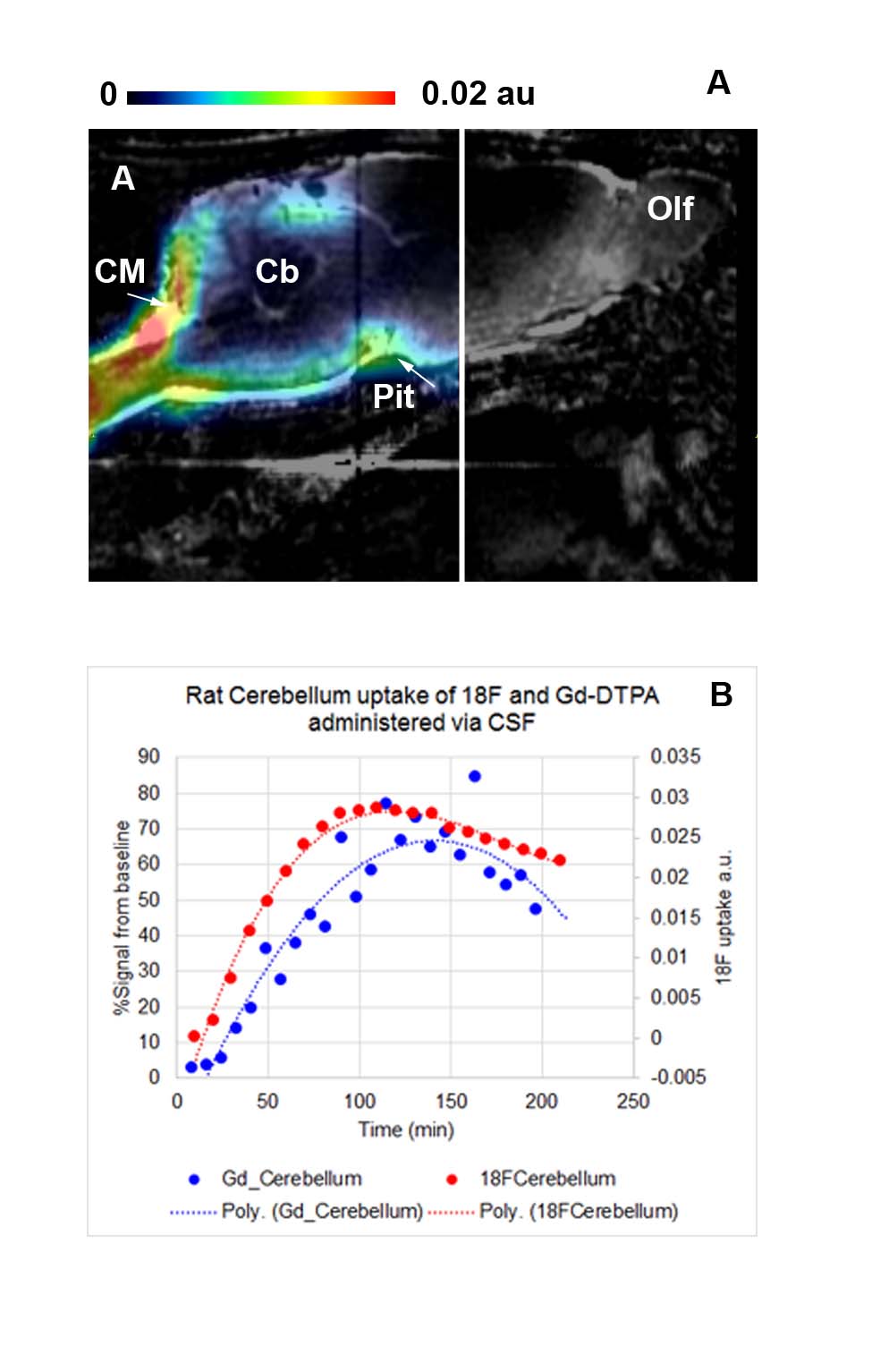
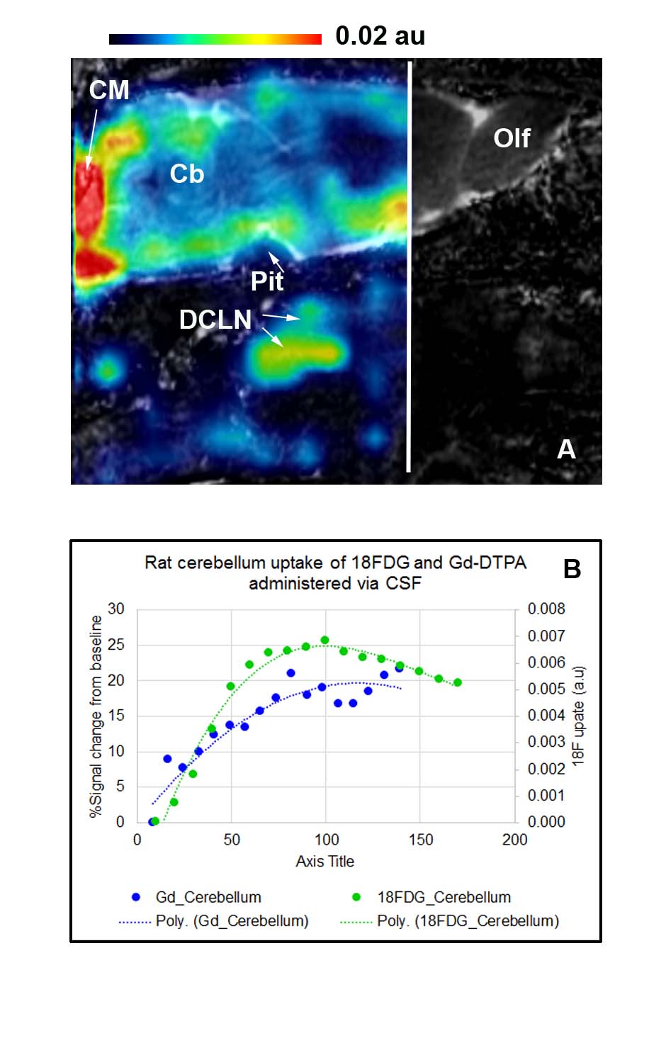
Fig. 1A shows the PET image of CSF transport of [18]F into rodent brain overlaid on the corresponding dynamic enhanced MRI at 1.5 hrs of circulation; and demonstrates high activity in the cisterna magna (input); along the peri-arterial channels and into the brain parenchyma. The corresponding time activity curves (TACs) for the [18]F tracer and Gd-DTPA show that [18]F parenchymal (cerebellum) glymphatic transport appears to be slightly faster when compared to Gd-DTPA (Fig. 1B). Fig. 2 shows a representative PET image of the brain-CSF distribution pattern of [18]FDG (FDG + metabolites) and Gd-DTPA after approximately 90min of tracer CSF circulation. Note that there is [18]FDG activity in the CSF spaces (cisterna magna (CM), in the cerebellum as well as other parts of the brain. In addition, there is uptake in areas associated with the deep cervical lymph nodes. The corresponding TACs for the metabolically active ([18]FDG) radiotracer and inert paramagnetic tracer (Gd-DTPA) show that [18[FDG (and metabolites) follows the same peri-vascular transport routes as Gd-DTPA, however, the uptake of the radiotracer is faster than Gd-DTPA (Fig. 2B).Discussion and interpretation
Using PET-MRI, we demonstrated that two different 18F-labeled radiotracers, 18F and 18FDG are transported from the CSF into the brain via peri-vascular routes and from these conduits into brain parenchyma; a transport pattern typical of glymphatic transport 1. To validate we co-administered Gd-DTPA and observed the characteristic glymphatic pathway distribution pattern including peri-vascular and brain parenchymal uptake as previously demonstrated5. Interestingly, [18]FDG tracer uptake was also observed in areas associated with the deep cervical lymph nodes suggesting drainage out of the brain. In contrast, no uptake of [18]F could be demonstrated in areas associated with deep cervical lymph nodes over the 2-3 hr experimental period which is suggestive of slow efflux of the tracer and/or minimal brain parenchymal uptake over time. We are in the process of validating these data using PET-CT and will be presenting these confirmatory data.Acknowledgements
Leducq foundation, AnonymousReferences
1. Nedergaard, M. Neuroscience. Garbage truck of the brain. Science 340, 1529-1530, (2013).
2. Iliff, J. J. et al. A paravascular pathway facilitates CSF flow through the brain parenchyma and the clearance of interstitial solutes, including amyloid beta. Science translational medicine 4, 147ra111, (2012).
3. Louveau, A. et al. Structural and functional features of central nervous system lymphatic vessels. Nature 523, 337-341, (2015).
4. Maramraju, S. H. et al. Small animal simultaneous PET/MRI: initial experiences in a 9.4 T microMRI. Phys Med Biol 56, 2459-2480, (2011).
5. Iliff, J. J. et al. Brain-wide pathway for waste clearance captured by contrast-enhanced MRI. J Clin Invest 123, 1299-1309, (2013).
Figures