3679
Evaluation of Diagnostic Accuracy of Gallium-68 DOTATATE PET-MRI Hybrid Imaging in Staging and Restaging Neuroendocrine Tumors1Department of Radiology, Stony Brook University Hospital, Stony Brook, NY, United States
Synopsis
The purpose of our study is to evaluate diagnostic accuracy of DOTATATE PET-MR hybrid imaging in detecting neuroendocrine tumor. We demonstrated Ga-68 PET-MRI has high accuracy in detection of NET in the abdomen and pelvis. Lesion characterization with combined MRI helps to distinguish benign lesions from malignant ones.
Purpose
In patients with NET, accurate diagnosis and staging allow the patient to receive the best treatment and outcome. Ga-68 DOTATATE PET-MR combines high somatostatin receptor affinity of DOTATATE and superior soft tissue delineation and characterization of MRI for neuroendocrine tumor (NET) evaluation. The purpose of the study is to assess diagnostic accuracy of DOTATATE PET-MR hybrid imaging in the detection of NET.METHOD AND MATERIALS
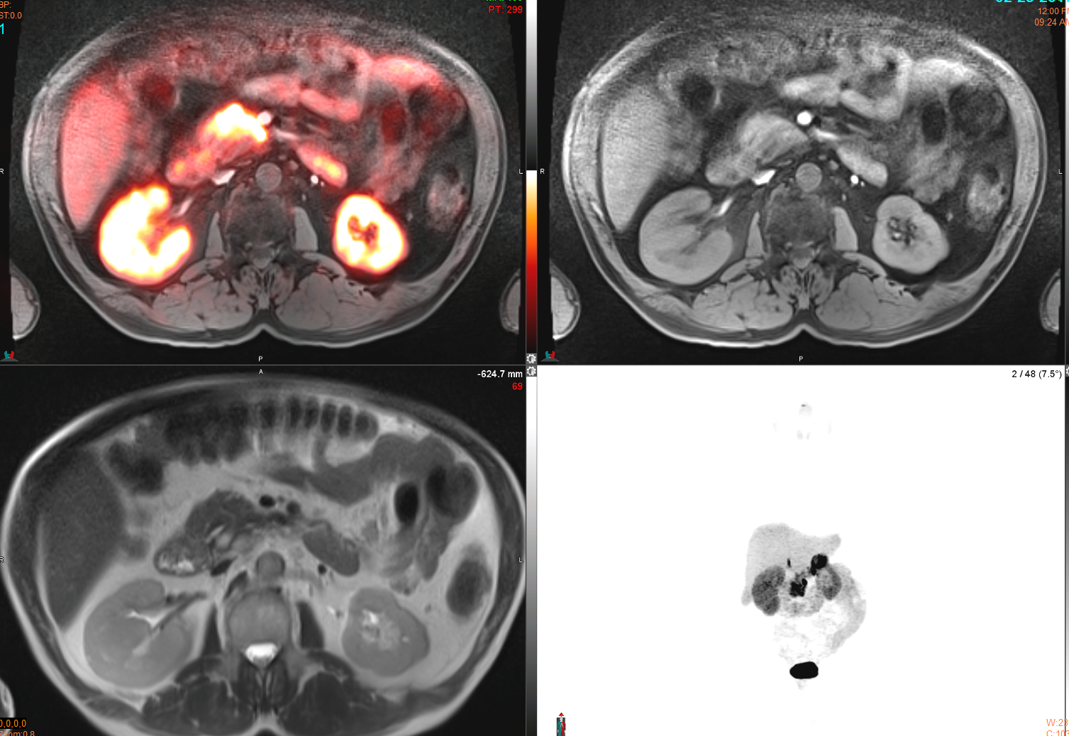
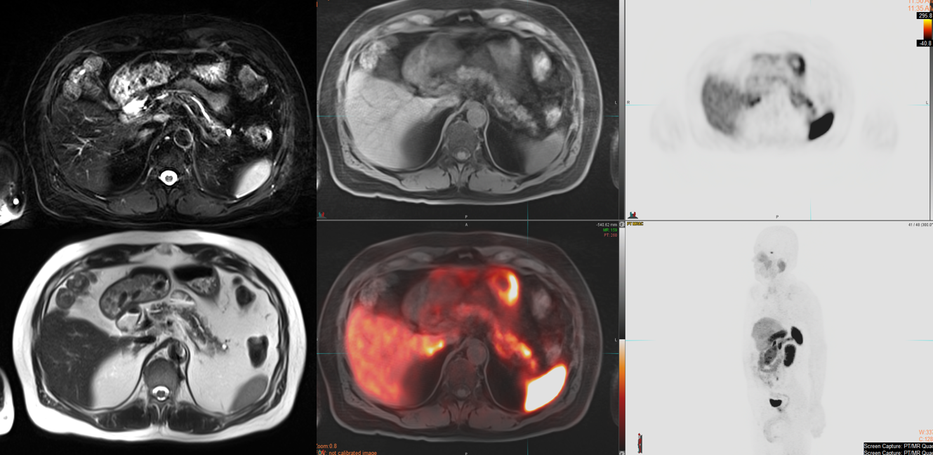
We retrospectively identified 11 patients (mean 62 yrs, 7 male, 4 female) with NET underwent combined Ga 68 DOTATATE PET-MRI for tumor staging and restaging between 2016 and 2017. 9 of 11 patients had metastatic carcinoid tumors and 2 of 11 patients had pancreatic neuroendocrine tumors. Each PET-MRI was evaluated in consensus by a nuclear radiologist and a body radiologist. Final diagnosis of positive NET was determined in 8 of 11 patients by histopathology, laboratory, and clinical follow up. Diagnosis of negative NET was confirmed in 3 of 11 patients.RESULTS
DOTATATE PET-MRI identified NET positive tumors in 8 of 8 patients (sensitivity = 100%) and exclude NET recurrence in 2 of 3 patients (Specificity = 67%). PET demonstrated 0 false negative result, and 1 false positive result (PPV=89%, NPV=100%). Patients had lesions in abdominal pelvic lymph nodes (N=6), liver (N=5), pancreas(N=2), bone (N=2), neck lymph nodes (N=2), and mediastinal lymph node (N=1), and cervical cuff (N=1). Oneprimary lesion was identified in the ileum. The false positive pancreatic lesion was confirmed to be sequela ofchronic pancreatitis. One patient had hypermetabolic pancreatic lesion falsely positive on DOTATATE, but MRI and pathology confirmed intraductal papillary mucinous tumor.CONCLUSION
Ga-68 DOTATATE PET-MRI demonstrates high accuracy in detection of NET in the abdomen and pelvis. Lesion characterization with combined MRI helps to distinguish benign lesions from malignant ones. Pathology may still be required in patients with equivocal or unusual findings.Acknowledgements
No acknowledgement found.References
1. Naswa N, Sharma P, Kumar A, Nazar AH, Kumar R, Chumber S, et al. Gallium-68-DOTA-NOC PET/CT of patients with gastroenteropancreatic neuroendocrine tumors: a prospective single-center study. AJR Am J Roentgenol. 2011;197(5):1221-8.
2. Maurer T, Gschwend JE, Rauscher I, Souvatzoglou M, Haller B, Weirich G, et al. Diagnostic Efficacy of (68)Gallium-PSMA Positron Emission Tomography Compared to Conventional Imaging for Lymph Node Staging of 130 Consecutive Patients with Intermediate to High Risk Prostate Cancer. J Urol. 2016;195(5):1436-43.
3. Oberg K. Gallium-68 somatostatin receptor PET/CT: is it time to replace (111)Indium DTPA octreotide for patients with neuroendocrine tumors? Endocrine. 2012;42(1):3-4.
4. Yang J, Kan Y, Ge BH, Yuan L, Li C, Zhao W. Diagnostic role of Gallium-68 DOTATOC and Gallium-68 DOTATATE PET in patients with neuroendocrine tumors: a meta-analysis. Acta radiologica. 2014;55(4):389-98.
5. Treglia G, Castaldi P, Rindi G, Giordano A, Rufini V. Diagnostic performance of Gallium-68 somatostatin receptor PET and PET/CT in patients with thoracic and gastroenteropancreatic neuroendocrine tumours: a meta-analysis. Endocrine. 2012;42(1):80-7.
Figures