3633
White Matter Differences in Parkinson’s Disease: a 7Tesla Tract-Based Spatial Statistics Study1Center for Magnetic Resonance Research, University of Minnesota, Minneapolis, MN, United States, 2Neurology, University of Minnesota, Minneapolis, MN, United States
Synopsis
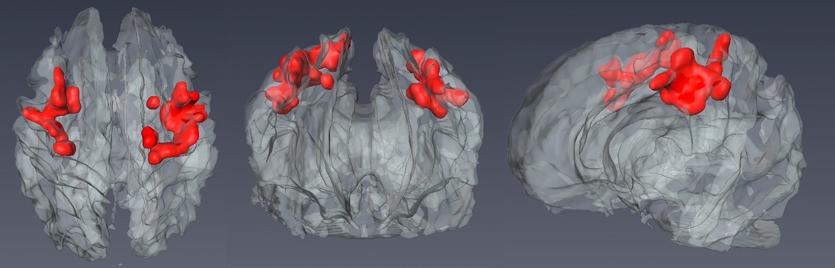
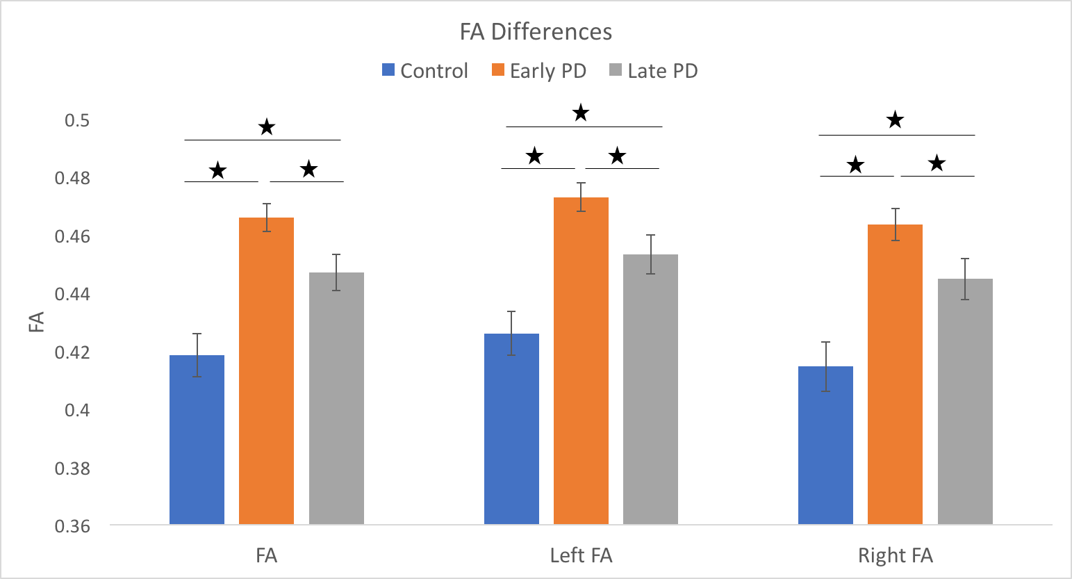
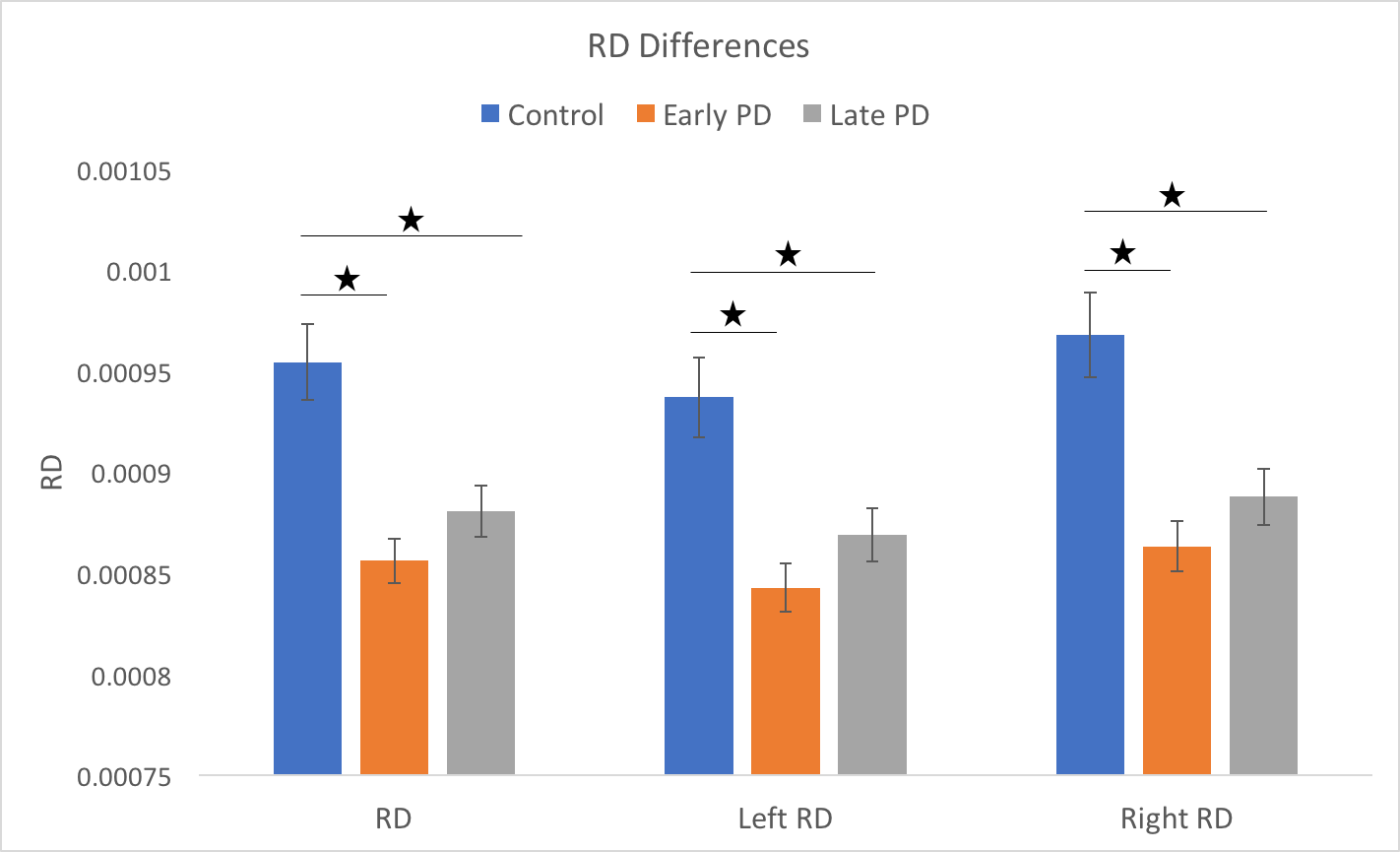
We used tract-based spatial statistics (TBBS) of 7Tesla fractional anisotropy (FA) measures to find differences between a cohort of early PD patients (N=30) and healthy controls (N=28). We examined the evolution of these differences by adding a PD cohort with longer disease duration (N=21). TBSS analysis revealed increased FA in the early PD group in the portions of the superior longitudinal fasciculus and the corticospinal tracts near M1, pre-motor and SMA. Subsequent analyses revealed increased FA in both PD groups compared to the control group. Radial diffusivity was significantly lower in the early and late PD groups compared to controls.
Introduction
Parkinson’s Disease (PD) is a neurodegenerative disorder characterized by a variety of motor symptoms that worsen over time. Several studies have used magnetic resonance imaging (MRI), especially diffusion MRI, to study white matter changes in PD1-6. In this study, we used tract-based spatial statistics7, at 7Tesla, to find differences between a cohort of people with early PD and healthy controls. Next, we examined the evolution of these differences cross-sectionally by comparing these white matter regions in a cohort with longer duration PD.Method
7T MRI data were acquired for thirty subjects with early PD (age = 64±7yr, 13F, disease duration = 2.1 ± 2.0yr) and eighteen healthy controls (age = 60 ± 10yr, 8F). Diffusion images with 1.25mm isotropic were corrected for distortion, motion, and eddy current effects. Fractional anisotropy (FA) maps were computed from the corrected data. Further, the FSL TBSS pipeline was used to compare FA values between the early PD and the control group. Significant voxels along the skeleton were isolated and back-projected onto each subject’s native space, then FA and radial diffusivity (RD) values were averaged over the clusters for further statistical analysis. Laterality of the results was assessed and radial diffusivity was computed and averaged over the same clusters for further study. Finally, a lPD group, made of twenty-one patients, with longer duration disease (age = 65 ± 11yr, 5F, disease duration = 8.2 ± 2.3yr) was introduced to study the evolution of the differences observed between the early PD and the control group. The FA and RD values were analyzed between the groups and post-hoc analyses were led within the early PD group.Results
A Chi-squared test revealed no significant differences in terms of the number of male and female participants between the groups (p=0.30). A T-test found no significant difference in age between the groups (p=0.06). TBSS analysis revealed bilateral clusters in the superior portions of the superior longitudinal fasciculus and the corticospinal tracts near motor (M1), premotor and supplementary motor (SMA) areas (Figure 1). Subsequent analyses revealed increased FA in the PD groups compared to the control group (p<0.0001 for early and p=0.009 for late patients)(Figure2). Additionally, FA values were significantly higher in the early PD group compared to the late PD group (p=0.02). Similar results were found when dividing into their left and right components (Figure2). Radial diffusivity was significantly lower in the early and late PD groups compared to controls (p=0.0003 and p=0.004, respectively)(Figure3).Discussion & Conclusion
Similar to our findings, one previous study has reported bilateral increases in FA in regions of the corticospinal tract originating from the M1, pre-motor and areas of people with mild to moderate PD4,6. These effects were interpreted to reflect a possible compensatory mechanism in early PD. Here, we show that these increases in FA are highest in early PD and that longer disease duration is associated with a significant reduction in this effect. Nonetheless, FA in the longer duration group was increased relative to controls. Finally, we show significantly lower RD in both patient groups compared to controls indicating a possible loss in myelin in these regions. These findings are in keeping with studies showing a significant increase in functional connectivity of corticofugal pathways in people with de novo (early untreated) PD8 suggesting that substantial compensatory changes in the morphology and function of corticofugal pathways occurs during prodromal and early stages of PD.Acknowledgements
This project was funded by NIH RO1-NS088679, NIH UL1TR000114, NIH R01-NS085188; P41 EB015894; P30 NS076408, MNDrive Fellowship to MP, and the University of Minnesota Udall center P50NS098573References
1. Georgiopoulos C, Warntjes M, Dizdar N, et al. Olfactory Impairment in Parkinson's Disease Studied with Diffusion Tensor and Magnetization Transfer Imaging. J Parkinsons Dis. 2017;7(2):301-311.
2. Ji L, Wang Y, Zhu D, Liu W, Shi J. White matter differences between multiple system atrophy (parkinsonian type) and Parkinson's disease: A diffusion tensor image study. Neuroscience. 2015;305:109-116.
3. Meijer FJ, van Rumund A, Tuladhar AM, et al. Conventional 3T brain MRI and diffusion tensor imaging in the diagnostic workup of early stage parkinsonism. Neuroradiology. 2015;57(7):655-669.
4. Mole JP, Subramanian L, Bracht T, Morris H, Metzler-Baddeley C, Linden DE. Increased fractional anisotropy in the motor tracts of Parkinson's disease suggests compensatory neuroplasticity or selective neurodegeneration. Eur Radiol. 2016;26(10):3327-3335.
5. Perea RD, Rada RC, Wilson J, et al. A Comparative White Matter Study with Parkinson's disease, Parkinson's Disease with Dementia and Alzheimer's Disease. J Alzheimers Dis Parkinsonism. 2013;3:123.
6. Wen MC, Heng HS, Ng SY, Tan LC, Chan LL, Tan EK. White matter microstructural characteristics in newly diagnosed Parkinson's disease: An unbiased whole-brain study. Sci Rep. 2016;6:35601.
7. Smith SM, Jenkinson M, Johansen-Berg H, et al. Tract-based spatial statistics: voxelwise analysis of multi-subject diffusion data. Neuroimage. 2006;31(4):1487-1505.
8. Kurani AS, Seidler RD, Burciu RG, et al. Subthalamic nucleus--sensorimotor cortex functional connectivity in de novo and moderate Parkinson's disease. Neurobiol Aging. 2015;36(1):462-469.
Figures