3618
Impaired microstructural integrity of the callosum forceps minor in type 2 diabetes mellitus affect bilateral frontal functional connectivity1Department of Radiology, the Affiliated Drum Tower Hospital of Nanjing University Medical School, Nanjing, China, 2Department of Endocrine, The Affiliated Drum Tower Hospital of Nanjing University Medical School, Nanjing, China
Synopsis
Type 2 diabetes mellitus (T2DM) is a risk factor for cognitive impairment. While its mechanism remains to be explored. In this study, using Automating Fiber-Tract Quantification (AFQ) analysis, we found that the fractional anisotropy (FA) in callosum forceps minor decreased in patients with T2DM, which indicated transverse white matter tracts connecting bilateral frontal cortex, was damaged. Meanwhile, functional connectivity between multiple brain regions within bilateral frontal cortex was decreased. The changes in the tract of callosum forceps minor might be the microstructural basis for functional changes of frontal cortex and help us understand the mechanism of T2DM related cognition decline.
Background
Type 2 diabetes mellitus (T2DM) has been reported to be associated with increased risk of cognitive impairment. Individuals with T2DM have 1.5-2.5 folds increased risk of dementia compared with those without diabetes[1]. However, the mechanisms underlying the effects of type 2 diabetes on brain structure and function remain controversial. The purpose of this study is to investigate the relationship of brain microstructure integrity and functional connectivity in patients with T2DM.Method
Participants including 30 patients with T2DM (12 males, age: 57.57±9.06 years, education degree: 11.8±3.23 years) and 30 non-diabetic control (NC) subjects were enrolled (14 males, age: 58.83±6.66 years, education degree: 10.67±2.54 years). Detailed neuropsychological assessment, clinical and biochemical information were collected. Meanwhile, diffusion tensor imaging (DTI) and Resting-State fMRI were acquired on a Philips Achieva 3.0T TX MR system equipped with an 8-channel phased array coil. Automating Fiber-Tract Quantification (AFQ) analysis was performed to measure tract profiles of DTI parameters for 20 white matter tracts[2]. The tract profiles of fractional anisotropy (FA) at 100 points along each of the 20 tracts were evaluated for the pointwise comparisons between two groups using independent sample T test analysis (p<0.05, FWE corrected). Regions of interest (ROIs) from the Human Brainnetome Atlas were used for resting-state functional connectivity (RSFC) analysis between two groups (p<0.05, NBS corrected)[3]Result
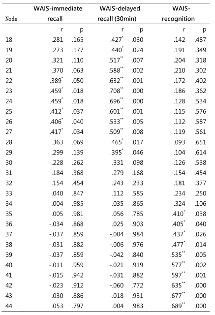
The MMSE score was significantly lower in patients with T2DM than NC group (28.9±0.95 vs. 28.22±1.28, p=0.045). Compared with the NC subjects, the T2DM patients showed regional FA reduction in the left part of the tract of callosum forceps minor, which is the only one with significant difference between groups in 20 of the examined fiber (Fig.1). Meanwhile, reduction of functional connectivity occurred mainly between the bilateral frontal cortex, including bilateral Orbital gyrus (OrG), bilateral Middle frontal gyrus (MFG), left Inferior frontal gyrus (IFG) and right Precentral gyrus (PrG) etc. (Fig.2). Further, we found that the FA values of multiple nodes in callosum forceps minor were positively correlated with Wechsler Abbreviated Scale of Intelligence (WASI) scores (Fig.3).Discussion
In patients with T2DM, fiber damage occurred in the callosum forceps minor, which includes transverse white matter tracts connecting the bilateral frontal cortex. RSFC analysis also confirmed the impaired functional connectivity of the bilateral frontal cortex in type 2 diabetic patients. Fiber damage was related to cognitive decline, especially the logical memory ability.Conclusion
The changes in the tract of callosum forceps minor might be the microstructural basis for functional changes of frontal lobe, which may add more information to understand the mechanism of cognition decline in patients with T2DM.Acknowledgements
This work was supported by the National Natural Science Foundation of China (81720108022, 91649116, 81571040, B.Zhang, 81471643, B.Zhu.); key medical talents of the Jiangsu province, the "13th Five-Year" health promotion project of the Jiangsu province (B.Z.2016-2020); the social development project of science and technology project in Jiangsu Province (BE2016605, BE201707); Jiangsu Provincial Key Medical Discipline (Laboratory) (ZDXKA2016020); the National and Provincial postdoctoral project (BE179 and 1501076A, BZ); the key project of Nanjing Health Bureau (ZKX14027, BZ); the Nanjing science and technology development program (NE179, B.Z.); and the project of the sixth peak of talented people (WSN-O50, BZ). National Key R&D Program of China (2016YFC0100100). The funders had no role in the study design, data collection and analysis, decision to publish, or preparation of the manuscript.References
[1]. Cheng, G., et al., Diabetes as a risk factor for dementia and mild cognitive impairment: a meta-analysis of longitudinal studies. Intern Med J, 2012. 42(5): p. 484-91.
[2]. Yeatman, J.D., et al., Tract profiles of white matter properties: automating fiber-tract quantification. PLoS One, 2012. 7(11): p. e49790.
[3]. Fan, L., et al., The Human Brainnetome Atlas: A New Brain Atlas Based on Connectional Architecture. Cereb Cortex, 2016. 26(8): p. 3508-26.
Figures
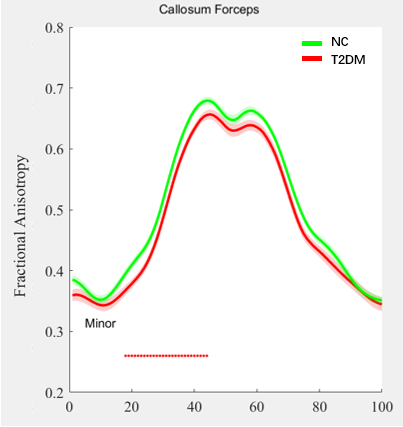
Fig.1: According to the AFQ analysis, T2DM patients showed FA reduction in 18-44 nodes of callosum forceps minor (p<0.05, FWE corrected).