3591
Relaxation-compensated multi-pool CEST signal at 7T MRI of WHO IV gliomas is dependent on the anatomic localization1Radiology, DKFZ, Heidelberg, Germany, 2Medical Physics in Radiology, DKFZ, Heidelberg, Germany, 3Neuropathology, University Hospital Heidelberg, Heidelberg, Germany, 4Neuroradiology, University Hospital Heidelberg, Heidelberg, Germany, 5Neurosurgery, University Hospital Heidelberg, Heidelberg, Germany, 6Neurology, University Hospital Heidelberg, Heidelberg, Germany, 7Max-Planck-Institut, Tübingen, Germany
Synopsis
As patients with WHO IV° gliomas are still having a dismal prognosis, further tumor characterization is needed. With prognosis and histopathological parameters being dependent on tumor localization, and Chemical-Exchange-Saturation-Transfer(CEST) MRI at 7T being one of the latest advances in tumor imaging, we have prospectively evaluated CEST signals in 21 patients. Amide CEST and ADC parameters are significantly different with regard to brain hemispheres and correlating. CEST NOE(Nuclear Overhauser-Effect) is not different with regard to brain hemispheres, but in case of contact to the subventricular zone, which is accompanied by worse prognosis. NOE is possibly showing complementary information to Amide CEST.
Introduction
WHO IV° gliomas are one of the most aggressive primary malignant brain tumors in adults. Several studies of clinical routine have already shown a dependency of prognosis and histopathological parameters with tumor localization, especially in case of contact to the subventricular zone[1, 2]. Further characterization of the tumor and of novel MRI techniques are needed, to understand the tumor’s behavior. With relaxation-compensated multi-pool Chemical-Exchange-Saturation-Transfer (CEST) MRI at 7T being one of the latest, highly promising MRI techniques[3], we have prospectively evaluated the localization dependency of 7T CEST MRI signal intensities and histopathological parameters in newly diagnosed untreated WHO IV° glioma patients.Methods
Twenty-one patients with newly diagnosed, untreated WHO IV° gliomas were prospectively included in this study and investigated at a 7T whole-body scanner (Siemens Healthcare, Erlangen, Germany). Mean CEST contrasts (Nuclear-Overhauser-Effect(NOE), Amide-Proton-Transfer(APT), and Downfield_NOE-suppressed APT(DNS)) are measured using the linear difference (LD) [4], and AREX metric (AREX) [3]. Maximum (MAX), maximum 90% (_90) and minimum 10% (_10) parameters are calculated. These CEST signal intensities, ADC parameters and histopathological parameters of the tumor volumes were evaluated with regard to extension, localization (brain hemisphere and brain lobe) and contact to the subventricular zone using non parametric Mann-Whitney test, ANOVA on ranks test, Receiver Operating Characeteristic (ROC) and Spearman correlation (p≤0.05).Results
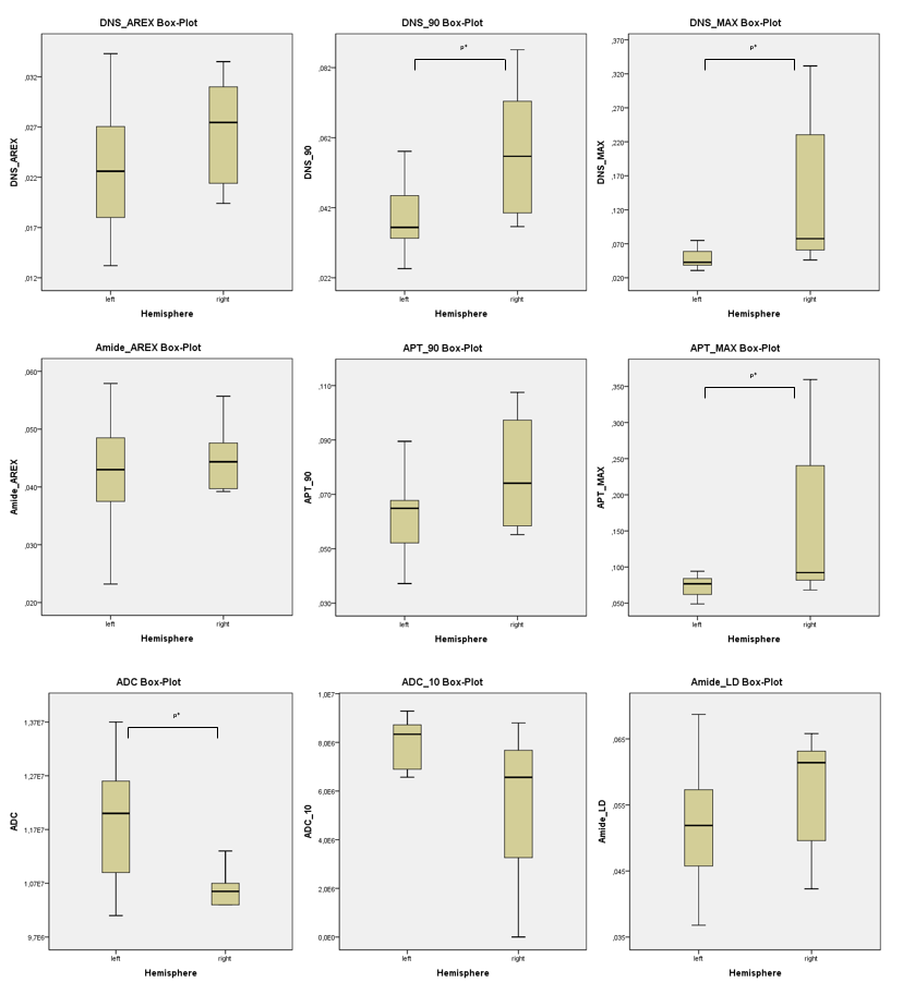
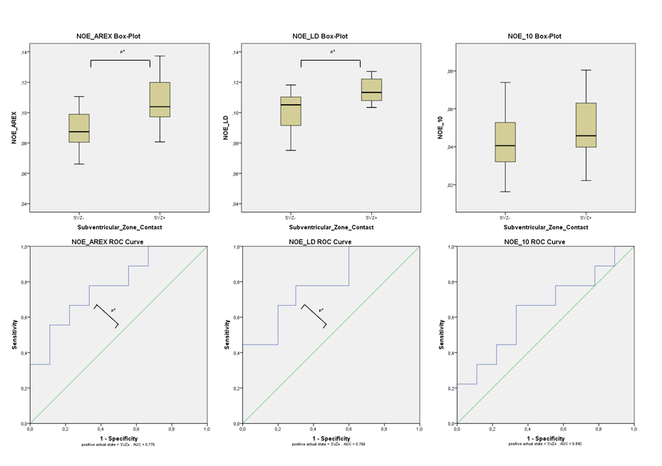
The Amide parameters APT_MAX, DNS_MAX and DNS_90 were significantly increased in right vs. left hemisphere gliomas (p=0.037, 0.024 and 0.007) (Figure 1). Mean ADC parameter were significantly decreased in right vs. left hemisphere glioblastomas (p=0.046) (Figure 1). Mean CEST NOE signal intensities did not differ significantly between both hemispheres, but were significantly increased in case of subventricular zone contact (NOE_AREX and NOE_LD), ROC curves had an area under the curve (AUC) of 0.778 and 0.789 (p=0.047 and 0.034) (Figure 2). The lobe localization and tumor extension did not have any significant effect on CEST and ADC parameters. CEST APT&DNS and ADC signal intensities did significantly correlate (-0.67 and -0.56) (p<0.01), but not CEST NOE and ADC parameters. Histopathological parameters were not significantly different with regard to different localizations.Discussion
This study is one of the first studies about prospectively evaluating localization dependency of signal intensities by means of relaxation-compensated multi-pool CEST MRI. Amide and NOE signals are showing different localization dependencies, which should be considered in future studies while interpreting NOE and amide CEST effects. Moreover, different localizations are characterized by different prognosis and histopathological parameters which might also reflect different signal intensitites in the investigated CEST contrasts. Nevertheless, confounding influence of spatial B0 and B1 inhomogeneities, despite performed corrections, should be taken into account.Conclusion
Relaxation-compensated multi-pool CEST MRI signals might be dependent on the anatomic localization. Amide CEST and ADC parameters are significantly correlating – NOE parameters do not correlate with ADC parameters and are solemly different with regard to subventricular zone contact, possibly showing complementary information to Amide CEST and ADC.Acknowledgements
No acknowledgement found.References
1. Adeberg, S., et al., A comparison of long-term survivors and short-term survivors with glioblastoma, subventricular zone involvement: a predictive factor for survival? Radiat Oncol, 2014. 9: p. 95.
2. Ellingson, B.M., et al., Anatomic localization of O6-methylguanine DNA methyltransferase (MGMT) promoter methylated and unmethylated tumors: a radiographic study in 358 de novo human glioblastomas. Neuroimage, 2012. 59(2): p. 908-16.
3. Zaiss, M., et al., Relaxation-compensated CEST-MRI of the human brain at 7 T: Unbiased insight into NOE and amide signal changes in human glioblastoma. NeuroImage, 2015. 112: p. 180-188.
4. Jones, C.K., et al., Nuclear Overhauser enhancement (NOE) imaging in the human brain at 7T. Neuroimage, 2013. 77: p. 114-24.
Figures