3588
A mannan-based probe for multimodal imaging of cancer and immune cells1IKEM, Prague, Czech Republic, 2UMCH, Prague, Czech Republic
Synopsis
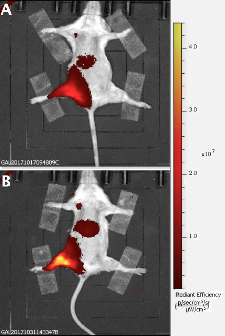
Presented mannan-based polymers have promising properties for tumor and metastasis imaging due to their biocompatibility, nanosize and specificity for the immune cells. In this study, two mannan-based polymers were tested by multimodal imaging (MRI and fluorescence). The polymers showed superior imaging properties compared to a commercially available contrast agent. FLI signal at the liver and higher signal at the injection site in the mouse with MN-Ox suggested slower elimination process due to addition of polyoxazoline chains in its structure. Both probes were visualized by MR and optical imaging modality at the injection sites and in the lymph nodes of the experimental mice suggesting their promising properties for cancer diagnosis.
Purpose
Mannan can be prepared in the form of nanoparticles allowing its use as a drug delivery carrier into solid tumors via Enhanced Permeability and Retention (EPR) effect. Moreover, due to its preferential uptake by immune cells (macrophages and dendritic cells) via DC-SIGN receptors1, mannan is preferably accumulated at the site of inflammation and in the lymph nodes. Therefore, mannan can serve as a multifunctional probe for diagnosis of tumors, metastasis and sentinel lymph nodes, as well as inflammation processes. In this study, mannan core was modified by a chelate containing gadolinium for magnetic resonance imaging (MRI) and by a near-infrared fluorescent dye for fluorescence imaging (FLI). Moreover, the probe was grafted with poly(2-methyl-2-oxazoline) for increasing of probability of accumulation in the tutor tissue. Mannan without (MN) and with oxazoline (MN-Ox) were characterised by MRI and FLI. Accumulation of the probes was assessed on the tumor-bearing mice.Methods
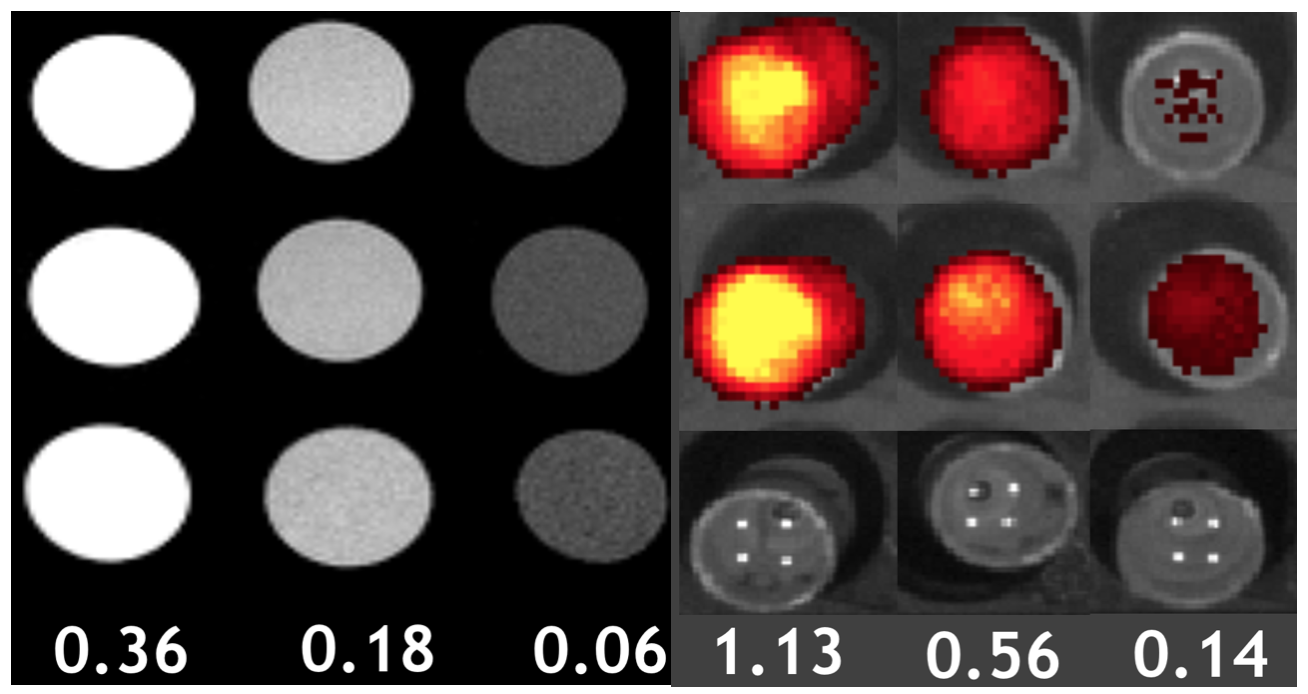
Results
Discussion
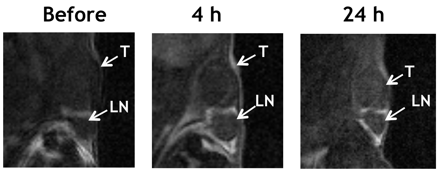
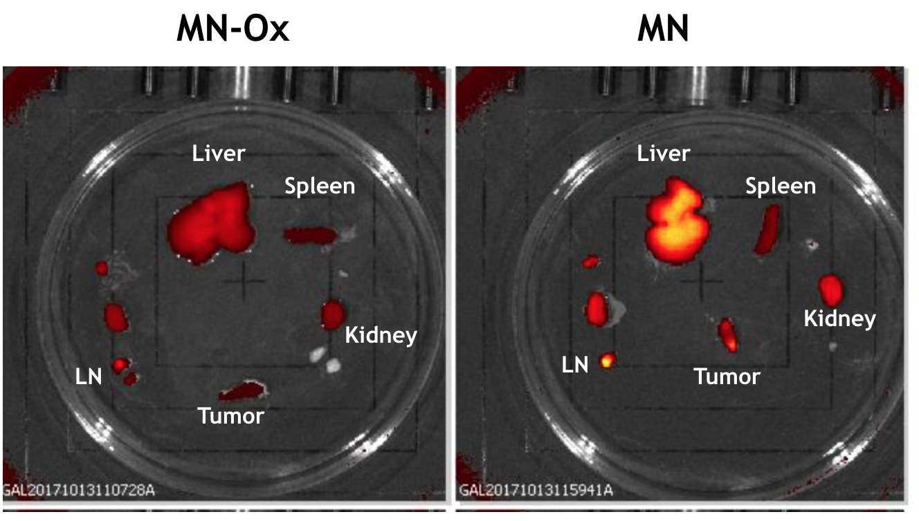
Accumulation of the mannan-based nanoprobes in the tumor tissue and the lymph nodes confirmed their targeting to immune cells and solid tumors. The mannan-based probes modified with were present at the injection sites for 7 days indicating slow biodegradation with the maximum of MRI and FLI signal at the first day following agents administration. Slower elimination rate of MN-Ox compared to MN was manifested by lower signal in the liver; however there was a lower FLI signal of MN-Ox in the tumor compared to MN probably due to refuse of oxazoline by the tumor-associated macrophages.Conclusion
Here, we present a novel multimodal sensitive mannan-based nanoprobes which target lymph nodes and tumor tissue. Easily modified chemical structure allows incorporation of drugs and makes this platform a suitable drug delivery system for theranostics of cancer, metastasis and inflammation.Acknowledgements
Supported by MH CR-DRO (Institute for Clinical and Experimental Medicine IKEM, IN00023001) and the Ministry of Health, Czech Republic (grant #15-25781A).References
[1] Cui Z. et al. Drug Dev Ind Pharm, 29(6), 2003Figures