3550
Accelerated real-time spiral MRI for high-resolution velum imaging using low rank and sparse decomposition and Chebyshev based off-resonance correction1Biomedical Engineering, University of Virginia, Charlottesville, VA, United States
Synopsis
In this study, an accelerated real-time spoiled spiral GRE sequence was developed for high-resolution velum imaging during speech to evaluate VPI. The low rank plus sparse decomposition was used to reconstruction highly undersampled (6x) dynamic image series. Chebysheve based off-resonance correction was used to reduce local blurring around velum after L+S reconstruction. The developed method generated high quality dynamic images with minimal temporal blurring and reduced blurring at 1.5 T, which can then be used to track velum movements for further analysis.
Introduction
Velopharyngeal insufficiency (VPI) is commonly seen in children who have a cleft palate repair. Due to no radiation exposure and good soft tissue contrast, real-time dynamic MRI acquired during speech can be used to accurately analyze the movement patterns of the tongue and velum to evaluate VPI with model-based muscle analysis 1,2. However, the slow speed of MRI poses challenge to satisfy the spatial and temporal resolution, coverage and SNR requirements simultaneously. An undersampled spiral GRE sequence combines the advantages of spiral scanning with parallel imaging and model-based temporal sparsity reconstruction to achieve maximal acceleration 3,4. Low rank and sparse decomposition (L+S) has been shown to perform better than original temporal sparsity, especially when global signal changes among frames 5. In addition, the air-tissue boundary around the velum causes non-linear off resonance that degrades image quality, especially at 3T. Chebyshev based off-resonance correction 6 can be combined with the L+S reconstruction to reduce blurring. In this study, we develop an imaging method with the aforementioned reconstruction framework.Methods
An interleaved spoiled spiral GRE sequence with linearly decreasing sampling density is used in this study. The fully sampled spiral k-space trajectory contains 18 interleaves with 3.6 ms readout per interleaf. 3 interleaves 120° apart from each other are used to reconstruct each frame, so that the undersampling rate is 6. Among frames the interleaves are rotated in a bit-reversed order to reduce temporal correlation. Reduced FOV (150 mm) is used with two spatial saturation pulses played every frame to improve spatial resolution. Other sequence parameters are: TR: 6.96 ms, TE: 0.78 ms, Flip Angle: 10°-20°. Simultaneous dual slice coverage with one mid sagittal and one oblique coronal slice is achieved by interleaving the TRs for the two slices and sharing the saturation bands. A low resolution field map is acquired with two single-shot spirals covering the center k-space. The spatial resolution is 1.2 mm and the effective temporal resolution is 30 fps when one slice is covered and 21 fps when two slices are covered.
In reconstruction, the dynamic image series is decomposed into low rank and sparse terms. Temporal differences are used as the sparsity constraint, since dynamic movements are limited to the tongue and velum regions. As multiple receiver coils are used in this study, in order to decompose on and solve for the combined image instead of individual channel images, the coil sensitivity is estimated from center k-space data during field map collection using ESPIRiT 7. To reduce local blurring around the velum, the Chebyshev based off-resonance correction method is used after L+S reconstruction by treating the resulting images as the 0th term and inverse gridding to obtain the un-acquired k-space data. A low resolution linear field map is used to improve the robustness of the Chebyshev method.
Experiments were performed on both Siemens Avanto 1.5 T and Prisma 3 T scanners on healthy volunteers. During the scan, the subjects are required to pronounce certain phrases such as “Bob ate apple pie”. The dynamic image series can then be used to segment the velum and calculate variables of interest for movement analysis.
Results and Discussions
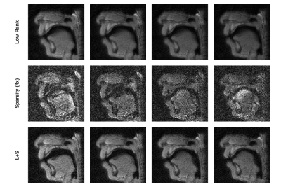
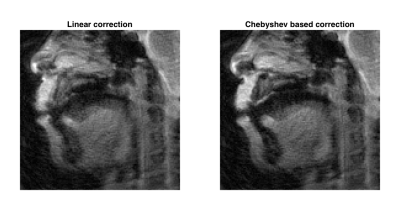
Fig. 1 shows the reconstructed dynamic image series obtained at 1.5 T and the corresponding decomposed low rank and sparse terms. As the images are acquired before the final steady state is reached, the low rank term captures the global contrast changes while the sparsity term focuses on the tongue movements. The final image series clearly shows the tongue movements with minimal temporal blurring and high SNR. Fig. 2 demonstrates the much improved off-resonance correction using the Chebyshev based method as compared with just correcting for the linear terms. However, due to much increased off resonance at 3 T, the image quality is significantly worse than at 1.5 T. As the muscle modeling also requires high resolution static velum images using 3D SPACE sequence, the study is preferred to run on 3 T due to higher SNR. Further improvements of the proposed dynamic MRI sequence at 3 T will be made including redesigning the k-space trajectory to reduce the readout time and optimizing the Chebyshev based correction method.Conclusion
An accelerated real-time spiral GRE sequence was implemented and tested in dynamic velum imaging on both 1.5 T and 3 T. The reconstruction framework with combined L+S and Chebyshev based off-resonance correction has showed success for highly undersampled data with minimal temporal blurring and reduced off resonance effects.Acknowledgements
No acknowledgement found.References
1. Pelland CM, Inouye JM, Feng X, et al. A new method for measuring soft palate deformations: MRI-based velum shape clustering. Boston, MA: 7th World Congress of Biomechanics. 2014
2. Pelland CM, Inouye JM, Feng X, et al. Use of dynamic MRI to quantify velopharyngeal contact length and differentiate velopharyngeal contact configurations among phonemes. Indianapolis, IN: 71st Annual Meeting of the American Cleft Palate-Craniofacial Association. 2014.
3. Kim YC, Narayanan SS, Nayak KS. Flexible retrospective selection of temporal resolution in real-time speech MRI using a golden-angle spiral view order. Magn Reson Med. 2011. 65(5):1365-1371.
4. Feng X, Inouye JM, Blemker S, et al. Assessment of velopharyngeal function with multi-planar high-resolution real-time spiral dynamic MRI. Salt Lake City, UT: 21st Annual Meeting of ISMRM. 2013.
5. Otazo R, Candes E, Sodickson DK. Low-rank plus sparse matrix decomposition for accelerated dynamic MRI with separation of background and dynamic components. Magn Reson Med. 2014. 73(3): 1125-1136.
6. Chen W, Sica CT, Meyer CH. Fast conjugate phase image reconstruction based on a Chebyshev approximation to correct for B0 field inhomogeneity and concomitant gradients. Magn Reson Med. 2008. 60(5): 1103-1111.
7. Uecker M, Lai P, Mruphy MJ, et al. ESPIRiT--an eigenvalue approach to autocalibrating parallel MRI: where SENSE meets GRAPPA. Magn Reson Med. 2014. 71(3): 990-1001.
Figures

