3491
Arterial spin labeling (ASL)-based radiomics features for predicting perfusion territory changes after carotid endarterectomy: a pilot study1Radiology, Peking Union Medical College Hospital, Chinese Academy of Medical Sciences, Peking Union Medical College, Beijing, China, 2. Huiying Medical Technology Co., Ltd, Beijing, China, 3GE Healthcare, MR Research China, Beijing, China, 4Peking Union Medical College Hospital, Beijing, China
Synopsis
To investigate whether radiomics can be apply to cerebrovascular disease and to develop features based on ASL for predicting perfusion territory change after carotid endarterectomy (CEA). A total of 1029 features were derived from ASL images, and 14 features were selected when comparing the differences between the two groups (select K best P<0.05). The selected features in difference are in agreement with visual inspection of collateral flow based on arterial transit artifact (ATA) on ASL.
Introduction
Radiomics is defined as extraction of high-throughput data from images, and provide large number of features for decision support1. So far radiomics is most well developed in oncology, and its use in diagnostic radiology has also drawn considerable attention. The perfusion territories of bilateral internal carotid arteries would alter in carotid stenosis patients, and restoration of perfusion territories to the normal distribution has been observed after CEA2. It was reported that effective collateral flow in carotid stenosis patients is associated with normalization of perfusion territories after CEA3. The purpose of this study was to investigate whether radiomics can be apply to cerebrovascular disease and to develop features based on ASL for predicting perfusion territory change after carotid endarterectomy (CEA).Methods
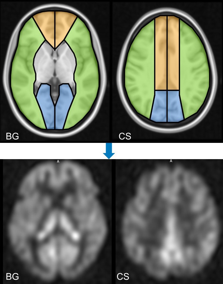
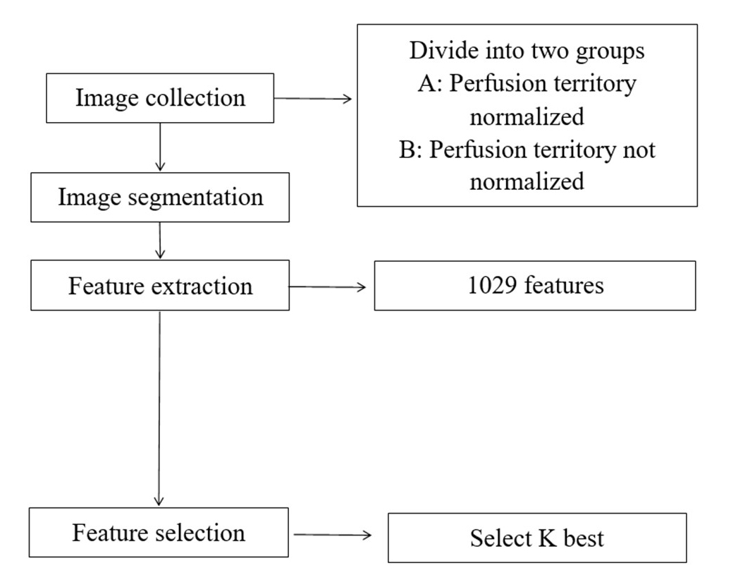
Twenty-one patients with severe carotid artery stenosis who underwent CEA were enrolled in this retrospective study. All these patients were imaged with ASL and vessel selective ASL before and after CEA. Common parameters of ASL and vessel selective ASL are as following: TR/TE 4886/10.5msec, labeling duration 1450msec, post labeling delay 2025ms, FOV 240mm× 240mm, matrix=512×512, slice thickness= 4 mm, slice gap= 0mm. Patients were divided into two groups according to whether perfusion territories revealed by vessel selective ASL were restored to normal after CEA2. Twelve regions of interest (ROIs) were defined on basal ganglia plane and centrum semiovale plane based on vascular territories: right/left anterior cerebral artery (ACA) region, right/left middle cerebral artery (MCA) region and right/left posterior cerebral artery (PCA), as shown in Fig.1. The ASL perfusion weighted images were normalized to the MNI space and masked with the ROI template. Regional perfusion features in these 12 ROIs were automatically extracted using select K best methods from ASL images before CEA. The work flow is illustrated in the flowchart in Fig.2.Results
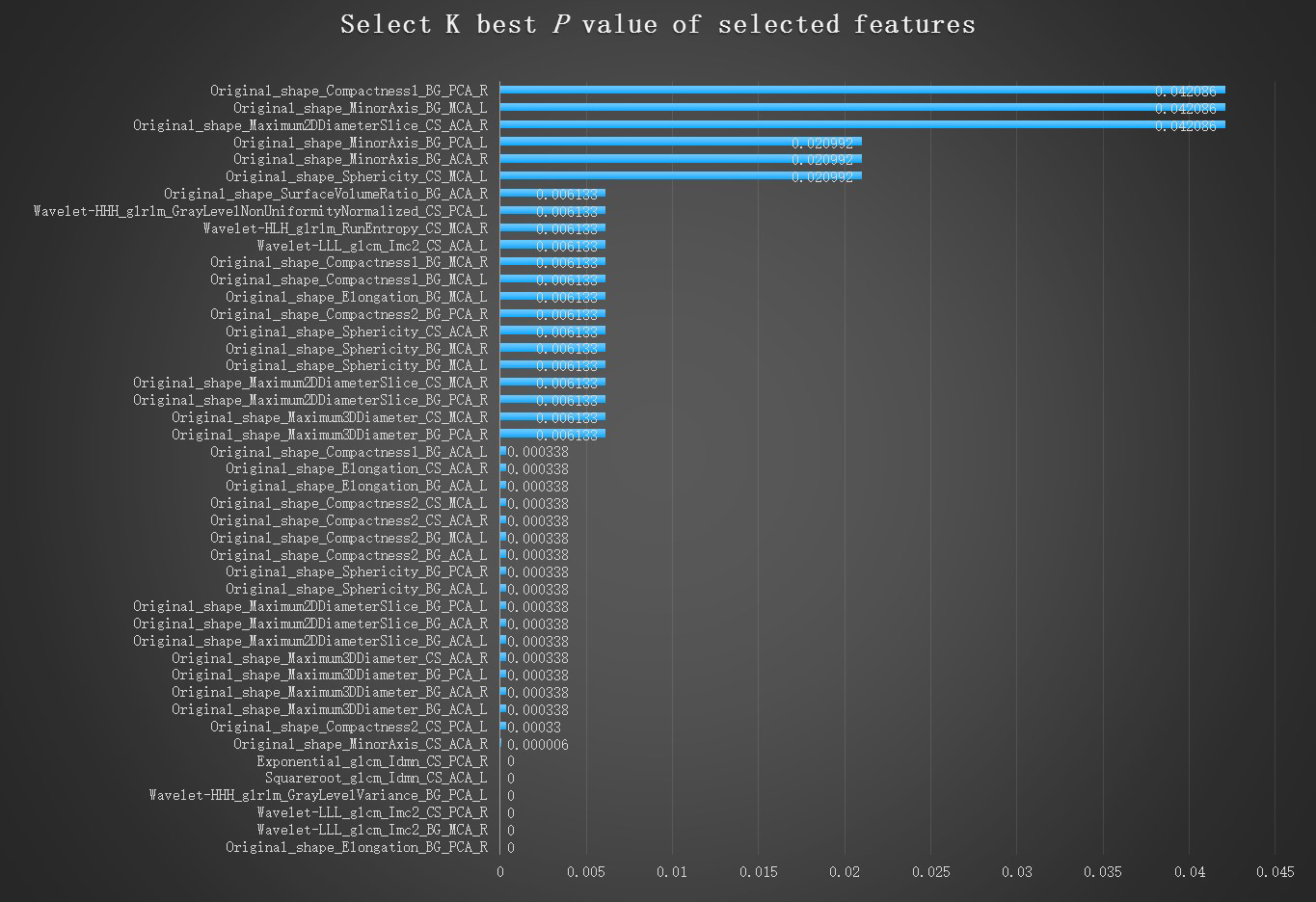
A total of 1029 features were derived from ASL images. Among these features, 14 features were selected when comparing the differences between the two groups (select K best P<0.05). ROI locations and P values of the selected features are shown in Fig.3. Four Original shape features: Maximum 3D Diameter, Maximum 2D Diameter Slice, Sphericity, and Compactness2 were the most commonly selected features, which were selected in six perfusion ROIs. Six features were found with extremely significant difference (P<0.000001) between the two different outcome groups. Among them, one is the original shape feature, elongation (defined as , λmajor and λminor are the lengths of the largest and second largest principal component axes); the rest five are higher order texture features: Gray Level Co-occurence Matrix (GLCM) and Gray Level Run Length Matrix (GLRLM) in different filter classes (i.e. wavelet-LLL, wavelet-HHH, squareroot and exponential).Conclusion and Discussion
In the previous study, collateral assessment was based on arterial transit artifact (ATA) on ASL perfusion weighted images, whose identification relies on objective visual inspection. ATA is defined as the bright serpiginous or spotted intravascular signal in ASL images. In this pilot study, radiomics method was investigated in the hope of deriving confined metrics that may predict collateral circulations. The shape feature elongation, with similar definition as ATA, was also selected in this radiomics method and showed significant difference in the two groups. The other texture features such as GLCM and GLRLM reflects the local spatial arrangement of intensities. The findings are in agreement with visual inspection that ATA appears as abnormal hyperintense in the relative low blood flow signal background. This pilot study indicated that radiomics method may be useful in predicting perfusion territory changes after CEA.Acknowledgements
We thank Mr. Guangqiang Geng for advice and assistance.References
1. Gillies RJ, Kinahan PE and Hricak H. Radiomics: Images Are More than Pictures, They Are Data. Radiology 2016; 278: 563-577. 2015/11/19. DOI: 10.1148/radiol.2015151169.
2. Van Laar PJ, Hendrikse J, Mali WP, et al. Altered flow territories after carotid stenting and carotid endarterectomy. Journal of vascular surgery 2007; 45: 1155-1161. 2007/06/05. DOI: 10.1016/j.jvs.2006.11.067.
Figures