3418
Quantification of Morphometry and Intensity Features of Intracranial Arteries from 3D TOF MRA: A Reproducibility Study1Electrical Engineering, University of Washington, Seattle, WA, United States, 2Radiology, University of Washington, Seattle, WA, United States, 3Surgery, University of Washington, Seattle, WA, United States
Synopsis
The aim is to evaluate the reproducibility of intracranial artery feature extraction (iCafe) technique for quantitative analysis of intracranial arteries from 3D time-of-flight (TOF) magnetic resonance angiography (MRA). Twenty-four patients with known intracranial artery stenosis were recruited and underwent two separate MRA scans within 2 weeks. Each dataset was processed blindly using iCafe. Eight morphometry and intensity features were acquired from each artery. The inter-scan reproducibility of iCafe was excellent with intra-class correlation coefficients between 0.92-0.98 and within-subject coefficients of variation between 3.2-8.6% across all features, showing iCafe is a reliable technique for intracranial artery feature quantification from TOF MRA.
INTRODUCTION
Intracranial atherosclerosis is one of the major causes for disability 1 and death 2 globally. As vascular structure and function are important factors for maintaining brain health, an accurate quantification of the whole intracranial vascular map could provide us with a method to evaluate cerebral vascular disease from the perspective of blood supply and vascular variations.
Intracranial artery feature extraction (iCafe) 3, a novel arterial feature extraction tool was recently developed to comprehensive quantify morphometry and intensity features for each artery and vascular group from 3D time-of-flight (TOF) magnetic resonance angiography (MRA).
Reliability of the extracted features are critical for further analysis of population data. Thus, reproducibility analysis is needed for two reasons. From the anatomical perspective, cerebral arteries are a complex network with substantial inter-individual variations 4. Whether iCafe could trace arteries accurately and quantify features consistently is unknown. From the perspective of imaging, it is well known that turbulence, slow or in-plane vessel flow will cause loss of intraluminal signal 5 and may cause differences between scans. Whether TOF MRA images could be reliably used for quantitative analysis of arteries is not clear.
In this study, we applied iCafe to a prospective cohort who underwent two separate MRA scans during separate scan sessions within two weeks. By evaluating representative features, we validated both the reproducibility of the iCafe technique and the consistency of TOF images.
METHODS
Patient studies
The dataset consisted of 24 clinical subjects with documented intracranial arterial stenosis. Subjects were scanned on a 3T Philips Ingenia Scanner using a standard head coil. Imaging parameters of TOF MRA were as follows: TR/TE = 14.7/3.5 ms, flip angle = 18°, in-plane resolution = 0.3 mm×0.3 mm, slice thickness = 1.4 mm, FOV = 190mm*190mm, matrix = 360*228, acquisition time = 118.92±20.18s. The time interval between the two scans was 10.17±2.97 days.
The study was approved by the Institutional Review Board and informed consent was obtained for all patients prior to enrollment.
Feature extraction
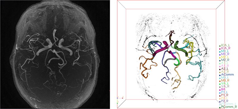
MRA images were resampled for isotropic resolution in 3D space. Field of view was aligned for each pair of images. Image intensities were normalized using the Nyul 6 method. Artery regions were then traced using an improved open-curve active contour model and labeled using a probability model in iCafe. Artery traces were visualized in Figure 1, and traces and labels could be corrected by user when errors occurred.
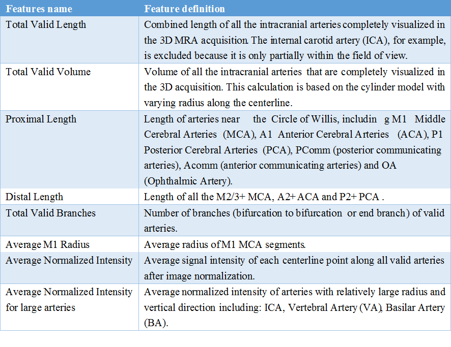
A total of 1456 morphometry and intensity features were extracted from each case. A representative subgroup of 8 features (listed in Table 1) was used to assess reproducibility.
Features reproducibility
MRA datasets were anonymized and randomized before being processed by the same experienced iCafe operator with an interval of at least one day for washout between processing the scan and rescan acquisitions. To test reproducibility, we calculated intra-class correlation coefficient (ICC) with 95% confidence interval (CI) and within-subject coefficient of variation (CV). Bland-Altman plot was used to visualize the differences. R studio (version 3.4.2) was used for the statistical analysis.
RESULTS
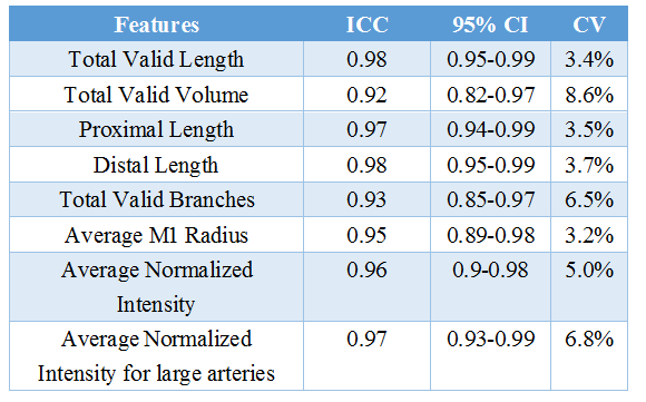
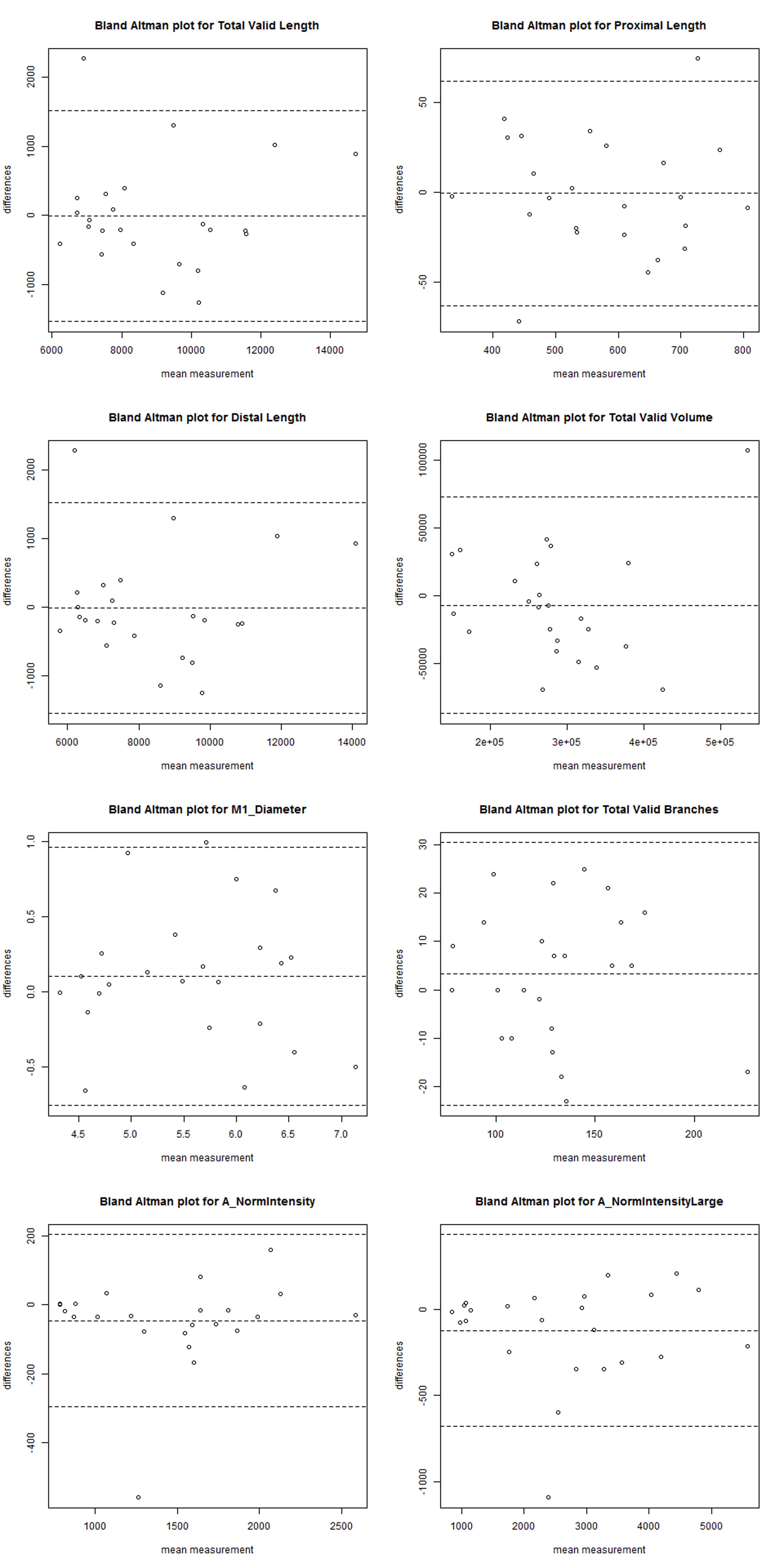
The 8 features showed excellent agreement, with ICCs from 0.92-0.98 and CVs between 3.2-8.6%, shown in Table 2. Bland-Altman plot was shown in Figure 2.
We found 1) all features showed excellent agreement in general; 2) the differences mainly occurred in short distal artery segments, as the “Length” reproducibility was better than “Total Valid Branches”; 3) the volume reproducibility was slightly less than that for length, as volume measurements incorporated length and radius, leading to increased variations; 4) length reproducibility of proximal and distal arteries was similarly excellent; 5) the relatively low intensity features were largely due to a single outlier case with large differences in intensity between acquisitions after normalization, possibly arising from large differences between the two scans or limitation of the normalization algorithm; 6) excellent average normalized intensity reproducibility of the large artery subgroup showed that loss of signal for in-plane vessel flow was not severe or was consistent; 7) mean intensity value from the subgroup of large arteries are reasonably higher.
DISCUSSION
Excellent reproducibility of the representative subset of features extracted by iCafe from the scan-rescan test shows it is a promising technique for morphometry and intensity feature extraction from TOF MRA images. As MRA is a frequently used sequence in clinical and research applications, this technique could provide a novel and reliable approach to evaluate intracranial vascular structure and blood flow in future investigational studies.CONCLUSION
TOF MRA is a reliable imaging sequence for quantitative analysis of intracranial arteries. iCafe can extract repeatable features for intracranial arteries from TOF MRA for future investigative quantitative analyses.Acknowledgements
This research is supported by grants from the National Institutes of Health (R01-NS083503-03, R01-NS092207-01A1, R01-HL103609-01A1) and Philips Healthcare.References
1. Murray CJL, Vos T, Lozano R, et al.: Disability-adjusted life years (DALYs) for 291 diseases and injuries in 21 regions, 1990-2010: a systematic analysis for the Global Burden of Disease Study 2010. Lancet 2012; 380:2197–2223.
2. Lozano R, Naghavi M, Foreman K, et al.: Global and regional mortality from 235 causes of death for 20 age groups in 1990 and 2010: a systematic analysis for the Global Burden of Disease Study 2010. Lancet 2012; 380:2095–2128.
3. Chen L, Mossa-Basha M, Balu N, et al.: Development of a quantitative intracranial vascular features extraction tool on 3DMRA using semiautomated open-curve active contour vessel tracing. Magn Reson Med 2017.
4. ALPERS BJ, BERRY RG, PADDISON RM: Anatomical Studies of the Circle of Willis in Normal Brain. Arch Neurol Psychiatry 1959; 81:409.
5. Carr JC (James C., Carroll TJ: Magnetic Resonance Angiography : Principles and Applications. Springer Science+Business Media, LLC; 2012.
6. Nyul LG, Udupa JK, Zhang X: New variants of a method of MRI scale standardization. IEEE Trans Med Imaging 2000; 19:143–150.
Figures