3400
Investigating the effect of B1 map inaccuracies on advanced pulse design1Danish Research Centre for Magnetic Resonance, Centre for Functional and Diagnostic Imaging and Research, Copenhagen University Hospital Hvidovre, Hvidovre, Denmark, 2Center for Magnetic Resonance, Department of Electrical Engineering, Technical University of Denmark, Lyngby, Denmark
Synopsis
B1 inhomogeneities at high field lead to undesired variation of contrast over the images. With advanced RF pulse design, the effect of B1 inhomogeneities on the excitation pattern can be restored. Bloch simulations in combination with advanced pulse design were performed to study the B1 mapping robustness. The results show that there is relatively high variation between four well established B1 mapping methods. From the results it is clear that next to acquisition speed and SNR, the robustness of B1 estimation is also an important factor if the B1 mapping is to be used for advanced RF pulse design.
Introduction
B1 inhomogeneities at high field lead to undesired variation of contrast over the images. With advanced gradient and RF pulse design, the effect of B1 inhomogeneities can be mitigated to an extent where the effective flip angle becomes more uniform, and a homogeneous contrast can be restored in the images. An outstanding problem in the field is the effect of B1 map inaccuracies on the resulting pulse design. There is currently no golden standard1, as most published methods suffer from either image distortions, limited sensitive dynamic range, motion sensitivity or long scanning times. In this abstract four established methods are assessed on robustness in RF pulse design.Methods
All experiments were performed on a Philips Achieva 7T MRI system (Philips Healthcare, Best, The Netherlands) equipped with a volume transit head coil and a 32channel receive coil array (Nova medical, Wilmington, MA, USA). All experiments were performed in accordance to the local ethical guidelines. Phantom experiments were performed using the fBIRN phantom2. A proof of principle was performed on one healthy volunteer. B1 maps were acquired using four established mapping techniques, which were actual flip angle (AFI), Bloch Siegert (BS), Dual Refocusing Echo Acquisition Mode (DREAM) and Saturated Dual Angle Method (SDAM)3-6.All data were acquired at an isotropic spatial resolution of 8 mm. Sequence parameters were as in Brink1, with exceptions of DREAM which had TR/TE,STE/TE,FID = 3.257/1.97/2.3 ms and SDAM which had TE = 5.95 ms.
Bloch simulations7 were performed in MATLAB (version 9.2.0, MathWorks, Natick, MA, USA) to study the effect of the different B1 mapping sequences on the excitation pattern. Iterative 2D spiral pulse design8 in the low-flip angle regime was performed to correct for B1 inhomogeneities. The error was quantified as the root mean squared (RMS) deviation from the target pattern. A cross comparison between methods was made where one method was used for optimization, and a second for verification in Bloch simulations. Additionally, experimental data was acquired with pulses designed with the four different sequences.
To assess the effect of motion between the B1 map acquisition, and the use of a numerically optimized pulse was estimated in simulation by moving the phantom, as well as by instructed motion of the volunteer for as far as the MR-coil allows. The error was again quantified as a RMS error in the excitation pattern.
Results
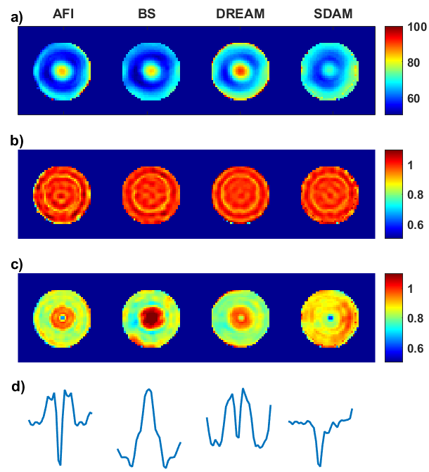
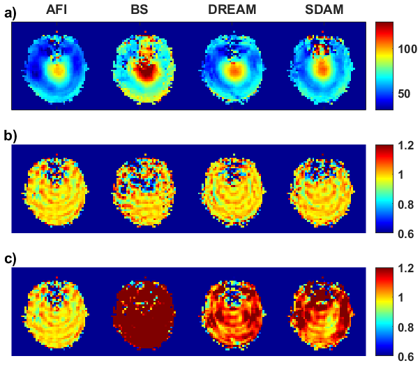
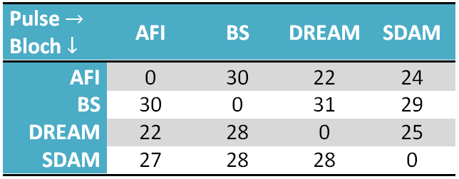
Phantom measurements (Fig. 1) show some differences in shape and scaling of the estimated B1 field. The BS-method shows a significant scaling difference, but also between the other methods clear differences are observed. Bloch simulations with a spiral excitation pulse show a high degree of uniformity in the predicted excitation pattern. Scanner results however show a slightly worse uniformity in the excitation pattern, especially in the central region (high B1 location). In vivo measurements (Fig. 2) show clear differences in shape and scaling of the estimated B1 field, Bloch simulations again predict a (false) high uniformity in the excitation pattern. Noise around to the nasal cavity due to worse B0 shimming is clearly visible in all methods, but most distinctive in the phase-based BS method. Table 1 shows that the RMS error results for the cross comparison are in the same range (22-30%) for all combinations, meaning that none of the B1 maps are interchangeable.
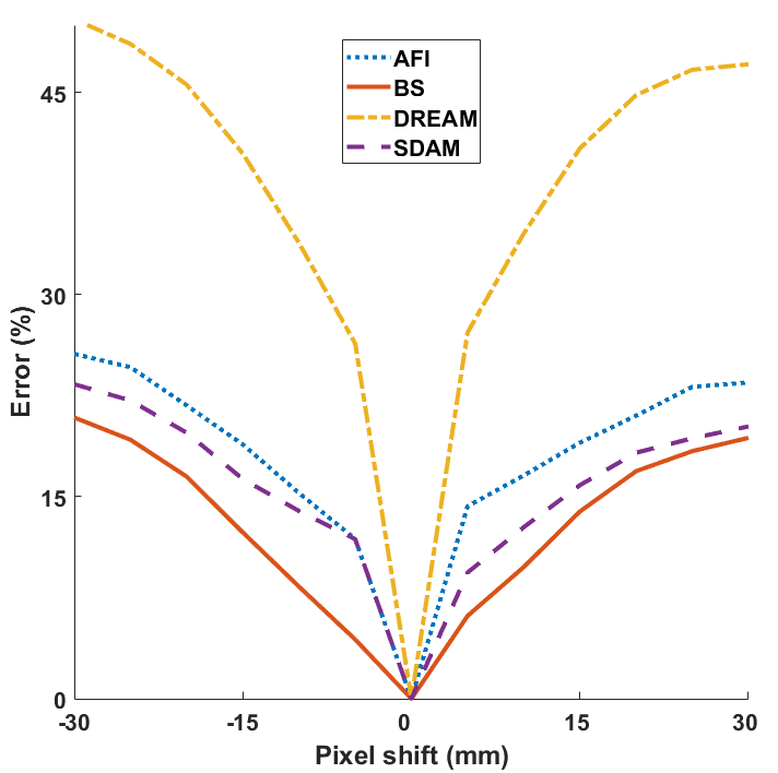
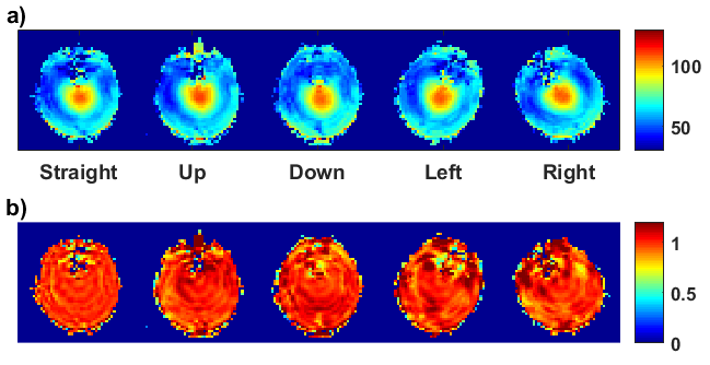
Simulated motion of the phantom (Fig. 3) in a diagonal direction results in errors for all methods, and especially for the DREAM method. More realistic motion of the volunteer (Fig. 4) shows a rotated B1 map in case of motion in the transverse plane, and a change around the nasal cavity in case of motion in the sagittal plane. Due to symmetry in the B1 field and the use of a small coil, there is not a large influence on the excitation pattern.
Discussion
The results show that there is relatively high variation between four well established B1 mapping methods. This could be due to several sources of systematic error, for example the SDAM method is most sensitive to motion, as a long TR and an EPI readout is used. Also the phase based BS method shows to be more sensitive to phase instabilities and motion as it relies on a subtraction technique. From these results it is clear that next to acquisition speed and SNR, the robustness of B1 estimation is also an important factor if the B1 mapping is to be used for advanced RF pulse design. Realistic motion inside a tight fitting receiver array does not seem to affect the result as such, however with less symmetric B1 fields, or coils that allow for more degrees of motion this should be re-assessed.Acknowledgements
This research is supported by the Danish Council for Independent Research [grant no. 6111-00349A].References
- Brink, W. M., Börnert, P., Nehrke, K., & Webb, A. G. (2014). Ventricular B1+ perturbation at 7 T–real effect or measurement artifact?. NMR in Biomedicine, 27(6), 617-620.
- Friedman L, Glover GH (2006) Report on a multicenter fMRI quality assurance protocol. J Magn Reson Imaging 23: 827–839
- Yarnykh, V. L. (2007). Actual flip‐angle imaging in the pulsed steady state: a method for rapid three‐dimensional mapping of the transmitted radiofrequency field. Magnetic resonance in Medicine, 57(1), 192-200.
- Sacolick, L. I., Wiesinger, F., Hancu, I. and Vogel, M. W. (2010), B1 mapping by Bloch-Siegert shift. Magn. Reson. Med., 63: 1315–1322.
- Nehrke, K., Versluis, M. J., Webb, A. and Börnert, P. (2014), Volumetric B1+ Mapping of the Brain at 7T using DREAM. Magn. Reson. Med., 71: 246–256.
- Cunningham, C. H., Pauly, J. M. and Nayak, K. S. (2006), Saturated double-angle method for rapid B1+ mapping. Magn. Reson. Med., 55: 1326–1333.
- Hargreaves, B. (2003), Bloch Simulator (MATLAB script).
- Yip, C. Y., Fessler, J. A. and Noll, D. C. (2005), Iterative RF pulse design for multidimensional, small-tip-angle selective excitation. Magn. Reson. Med., 54: 908–917.
Figures