3387
Parcellated shimming for brain imaging with 3D EPI at 7T1Sir Peter Mansfield Imaging Centre, School of Physics and Astronomy, University of Nottingham, Nottingham, United Kingdom
Synopsis
A novel acquisition strategy to improve the overall B0 field homogeneity by utilising 2D RF selection with acceleration via parallel transmission in conjunction with parcellated sub-volume shimming is proposed. The method has been demonstrated for brain imaging using a 3D EPI sequence at 7T.
Introduction
We implement a novel acquisition strategy to improve the overall B0 field homogeneity by utilising 2D RF selection with acceleration via parallel transmission in conjunction with local 2nd order volume shimming. Sequences with long echo-trains such as EPI1 suffer geometric distortion2 due to B0 inhomogeneity, along with TE-dependent signal drop-out. These effects impact upon fMRI experiments carried out at 7T3. In most studies, the effects of B0 inhomogeneity are ameliorated by shimming, which is performed so as to reduce the field variation over the whole brain volume that is being imaged 4. Dynamic shimming on a slice-by-slice basis has been shown to improve shimming efficiency5,6. Previous work based on simulations has shown that further gains in shimming efficiency can obtained by dividing the global volume into compact parcellated sub-volumes rather than slices7. Efficient implementation of this approach requires the use of two or three-dimensional selective excitation of sub-volumes, which is now feasible through the use of parallel transmit technology8-10.Method
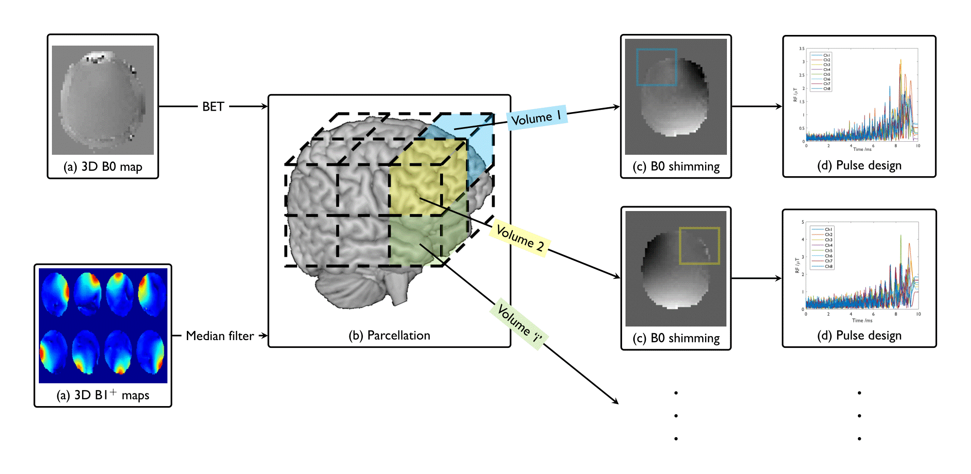
Experiments were performed on 4 volunteers using a multi-transmit-equipped Philips 7T Achieva scanner (Best, Netherlands) fitted with an 8-channel transmit/32-channel receive head coil (Nova Medical Inc, Wilmington). The experimental workflow is described in Figure 1. 3D B0-field maps with (4x4x8)mm3 resolution were acquired using a dual gradient echo sequence (FA=8°, TR=3.8ms,TE=1.52ms,ΔTE=1ms). B1 field maps for the 8-transmit elements were calculated from a single B1 map acquired in Tx quadrature mode (AFI11, FA=60°, TR1/TR2=30ms/150ms) and eight individual channel FFEs (FA=15°, TR=7.1ms)12.
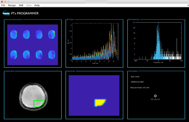
All B0 field shimming; RF and gradient waveform design was performed online using in-house software (Figure 2) written in Matlab (Natwick, USA) and Java. Brain extraction (BET13) was performed on B0 field maps. Relative B1 maps were median-filtered and appropriately thresholded. 12 parcellated volumes (6 superior, 6 inferior) were interactively selected to cover the whole brain volume. Design of RF pulses for exciting each sub-volume took 4 minutes. To limit total scan time, only 6 out of 12 sub-volumes were collected foreach volunteer.
Shim currents were calculated separately for each of the sub-volumes using a current-constrained minimization solver and B0 field maps were reacquired after shimming in each case for validation. A predesigned spiral 2D-k-space trajectory (duration=10ms, acceleration=3.5) was used for all inner-volume imaging with gradient pre-emphasis10,16. Multichannel pulses were designed for each of the sub-volumes using a small-tip-angle regularised approach14.
3D T1w-EPI was used for all imaging with a whole brain field-of-view with 3mm isotropic resolution (scan duration 11s, TE=27ms, TR=100ms, FA=10°), matrix size (196x196x120). To demonstrate proof-of-principle, each sub-volume was imaged separately to avoid the effects of eddy-currents from shim coil current switching. Zoomed imaging of sub-volumes was also demonstrated with 1.5mm2 in-plane resolution with matrix size (98x98x120) using half-Fourier acquisition (0.85-factor).
Results
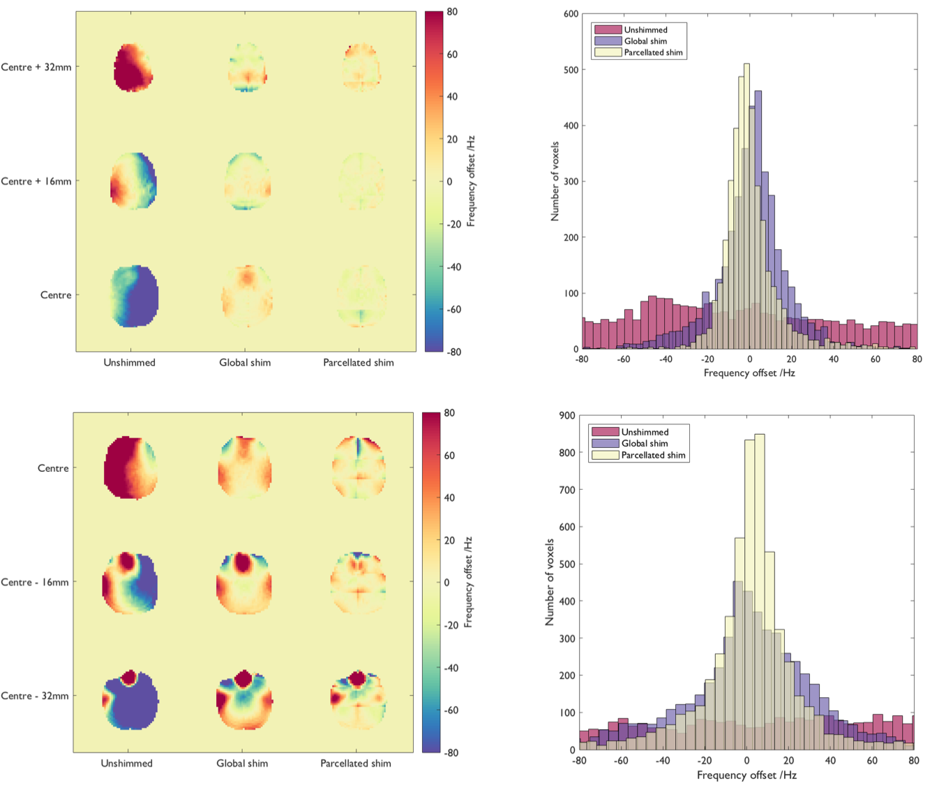
The standard deviation of the B0 field inhomogeneity over the combined superior sub-volumes was reduced by 22% (volunteer 1) and 23% (volunteer 2) through use of
parcellated B0 shimming as compared to global B0 shimming. Similarly, the standard deviation of the B0-field inhomogeneity over the combined inferior sub-volumes was reduced by 26% (volunteer 3) and 32% (volunteer 4).
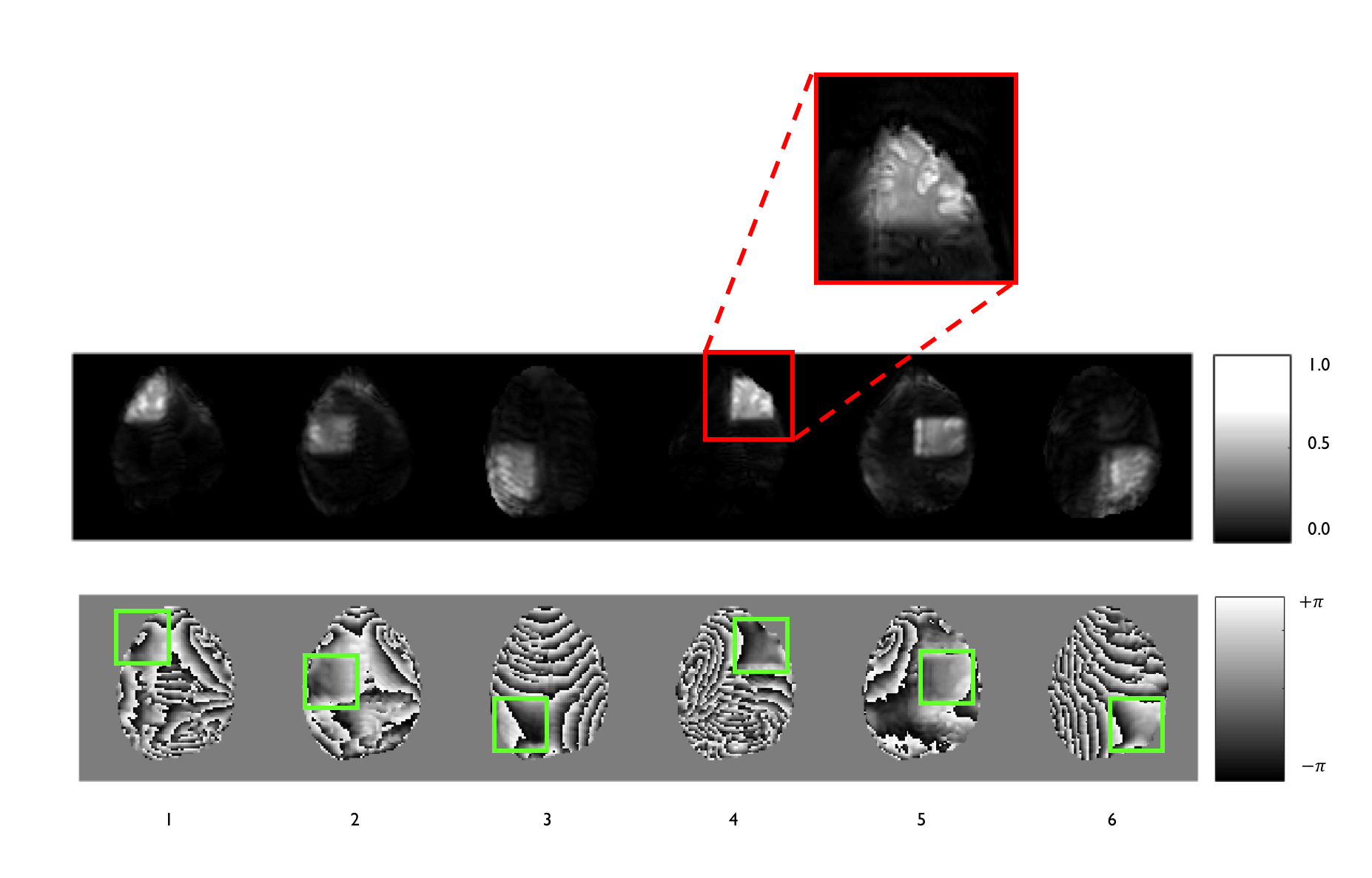
Figure 3 shows the 3D EPI magnitude and
phase for volunteer 1. The signal is localised within each desired sub-volume, and the low spatial variation of field over the selected-volumes is evident. Figure 4 displays the associated mean-centred histograms and
reconstructed frequency offset maps for volunteers 1 and 3.
Discussion
We have demonstrated a novel acquisition strategy to improve the efficacy of B0 shimming by using 2D-selective excitation pulses accelerated by parallel transmission to image compact parcellated sub-volumes. This approach improves the shimming performance because the field inhomogeneity over a set of compact sub-volumes can be better represented using a small number of spherical harmonic terms than is possible for the whole brain or a set of slices7. Full realisation of the benefits of this approach will require dynamic cycling of each excitation and sub-volume shim across TRs, rather than the separate sub-volume acquisitions demonstrated here. For this, the eddy-currents induced by fast switching of higher-order shim coils needs to be considered15,16.
Use of 2D-selective excitation produces a columnar rather than cuboidal sub-volume, so an optimal cycling of sub-volume excitations should be considered. The use of 3D-selective pulses17, with recent work18 showing that k-space trajectory optimisation can drastically reduce pulse durations (below 10ms) and would allow excitation of non-cuboidal sub-volumes. A practical limitation for this work was the pulse design time for each sub-volume, which limited the number of sub-volumes that could be acquired with the volunteer in situ, indicating the requirement for a parallelised design in the future.
Acknowledgements
This work was supported by funding from the Engineering and Physical Sciences Research Council (EPSRC) and Medical Research Council (MRC) [grant number EP/L016052/1]. The authors acknowledge the support of Philips Healthcare Clinical Services. The authors thank Shaihan Malik for support and valuable discussions.References
[1] Mansfield, P. Multi-planar image formation using NMR spin echoes. J. Phys. C: Solid State Physics. 10: L55-L58.
[2] Jezzard, P. & Balaban, R.S. Correction for Geometric Distortion in Echo Planar Images from B0 Field Variations. Magn. Reson. Med. 1995;34(1):65-73.
[3] Jezzard P. & Clare, S. Sources of distortion in function MRI data. Human Brain Mapping. 1999;8(2-3):80-85.
[4] Stockmann, J.P. & Wald, L.L. In Vivo B0 Field Shimming for MRI at 7T. Neuroimage. 2017; Article in Press.
[5] Morell, G. & Spielman, D. Dynamic shimming for multi-slice magnetic resonance imaging. Magn. Reson. Med. 1997;38(3):477-483.
[6] Sengupta, S. et al. Dynamic B0 shimming at 7T. Magn. Reson. Med. 2011;29(4):483-496.
[7] Poole, M. & Bowtell, R. Volume Parcellation for improved dynamic shimming. Magn. Reson. Mater. Phy. 2008;21(1):31-40.
[8] Katscher, U. et al. Transmit SENSE. Magn. Reson. Med. 2003;49(1):144-150.
[9] Schneider, J.T. et al. Inner-Volume Imaging In Vivo Using Three-Dimensional Parallel Spatially Selective Excitation. Magn. Reson. Med. 2013;69:1367-1378.
[10] Malik, S. & Hajnal, J. Phase Relaxed Localized Excitation Pulses for Inner Volume Fast Spin Echo Imaging. Magn. Reson. Med. 2016;76(3):848-61.
[11] Yarnykh, VL. Actual flip-angle imaging in the pulsed steady state: a method for rapid three-dimensional mapping of the transmitted radiofrequency field. Magn. Reson. Med. 2007;57(1):192-200.
[12] Van de Moortele, P-F. et al. (2007): In Proc. 15th ISMRM (Berlin). Abstract number 1676.
[13] Smith, S.M. Fast robust automated brain extraction. Human Brain Mapping. 2002;17(3): 143-155.
[14] Grissom, W. et al. Spatial domain method for the design of RF pulses in multicoil parallel excitation. Magn. Reson. Med. 2006;56:620-629.
[15] Fillmer, A. et al. Fast iterative pre-emphasis calibration method enabling third-order dynamic shim updated fMRI. Magn. Reson. Med. 2016;75:1119-1131.
[16] Çavuşoğlu, M. et al. VERSE-guided parallel RF excitations using dynamic field correction. NMR in Biomedicine. 2017;30(6):e3697.
[17] Wong, S.T.S. & Roos, M. A strategy for Sampling on a Sphere Applied to 3D Selective RF Pulse Design. Magn. Reson. Med. 1994;32:778-784.
[18] Davids, M. et al. Fast three-dimensional inner volume excitations using parallel transmission and optimized k-space trajectories. Magn. Reson. Med. 2016;76:1170-1182.
Figures