3385
Artificial Neural Network for Suppression of Metal Artifacts with Slice Encoding for Metal Artifact Correction (SEMAC) MRI1Magnetic Resonance Imaging Laboratory, Department of Bio and Brain Engineering, Korea Advanced Institute of Science and Technology, Daejeon, Republic of Korea, 2Department of Radiology, Seoul National University Hospital, Seoul, Republic of Korea
Synopsis
We present a new method of artificial neural network (ANN) to suppress metal artifacts in MR Imaging with Slice Encoding for Metal Artifact Correction (SEMAC). Seven titanium‑embedded phantoms were imaged using different SEMAC factors. The acquired data with low and high SEMAC factors were separated into input and label images, respectively, for training. The trained model was tested on separate phantoms. Metal artifacts in low SEMAC factors could be further suppressed visually and quantitatively using the implemented ANN, with the performance being comparable to that of label images. The proposed method reduces scan time necessary for high‑quality SEMAC imaging.
Introduction
Even though anatomical MR imaging for diagnostic purposes has become more readily available, patients with the necessity of imaging body regions with metal transplants, such as knee replacement and hip prosthesis, still do not benefit from the increased availability due to metal artifacts. There have been many trials to reduce metal artifacts for MR imaging. Lu et al. introduced Slice Encoding for Metal Artifact Correction in MRI (SEMAC) to suppress metal artifacts using extra z-phase encoding steps (SEMAC factor) combining with the View-Angle-Tilting (VAT) technique developed by Cho, et al.1,2. However, prolonged scan time for higher SEMAC factor imaging still remains as the technique’s inherent problem. In our study, we introduce artificial neural network to accelerate SEMAC imaging to suppress metal artifacts in a shorter scan time with comparable image quality.Methods
Multilayer Perceptron (MLP) is one of the most commonly used artificial neural network architectures, through which a fully connected hidden layer maps input values into output values. While Convolutional Neural Network (CNN) has become a dominating trend for machine learning, MLP has still proven to be useful for suppressing artifacts in MRI data 3.
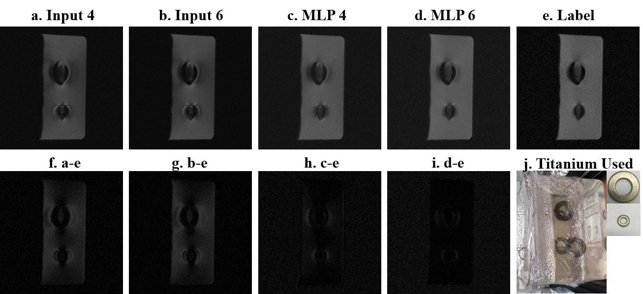
In SEMAC technique, extra z-phase encoding steps are implemented for each slice acquisition, from which signals from other z positions (partitions) are acquired and resolved back to their physical locations as a post processing. The number of extra‑encoding steps are called SEMAC factor, and ideally, increasing the factor enhances metal artifact suppression. In this work, low and high SEMAC factor images were categorized into input and ground truth, respectively, and were trained with MLP, through which output images were produced and compared with label images.
Input data were partitions of SEMAC factors 6 and 4, and ground truth data were those of SEMAC factor 12. Partitions of SEMAC factors 4 and 6 were retrospectively acquired from partitions of SEMAC factor 12. We compared partitions of SEMAC factor 6 from the retrospective sampling and those from real acquisition and found no significant difference between the two.
All data acquisitions were performed on a SIEMENS Skyra 3.0 T scanner (Siemens Medical Solutions, Erlangen, Germany). Total of 7 phantoms with different titanium metals embedded in 4% agarose were scanned along multiple directions to collect total 12 image sets. For both SEMAC factors of 6 and 12, proton-density weighted images (PD) were acquired, and the imaging parameters were: TR/TE = 3500.0/34.0 ms, FOV = 180 x 180 mm2, number of slices = 16/18/20 (depending on titanium), slice thickness = 3.0mm, matrix size = 256 x 256, flip angle = 140, and acquisition bandwidth = 610Hz/Px. Two out of total datasets were used for testing and the remaining ten datasets were used for training. This procedure was repeated by changing the two test datasets so that one dataset from every phantom was tested. Normalized root mean square error from the ground truth (NRMSE) was quantified for the analysis of the tested images. Wilcoxon signed rank test was performed to compare NRMSE and p value less than 0.05 was considered statistically significant.
Results
MLP showed smaller NRMSE than that of the input partitions, a trend observable regardless of SEMAC factor. The reduction in NRMSE using MLP was statistically significant for both input SEMAC factors 4 and 6 (p < 0.01) (Table 1). Figure 1 shows metal artifact suppression on a PD image of two titanium washers. The dark signal loss around the M10 (big) washer and signal pileup around M4 (small) washer were clearly corrected. Simple visual comparison of g-i suggests that artifact removal through MLP was comparable to the amount of correction in the label image, and that the suppressions were visibly significant for both factors 4 and 6.Discussion
The scan time for SEMAC factor 12 (12.7 min in this study) was double the scan time for SEMAC factor 6. Linear increase of scanning time with the SEMAC factor is a major problem of SEMAC imaging. Our study introduces a new effective way to reduce the scan time necessary for imaging with high SEMAC factor while maintaining the comparable quality of metal artifact suppression. Reducing scan time for SEMAC imaging is also related to a safety issue for imaging metal implants in vivo. When imaging patients with metal implants, prolonged scan time can induce tissue heating 4, and such risk can be also reduced through our method. While multiple phantoms are generated and tested in this study, the trained model can be additionally trained with in vivo data for real application in clinics, which warrants further investigation.Acknowledgements
No acknowledgement found.References
1. Lu, W., et al., SEMAC: Slice Encoding for Metal Artifact Correction in MRI. Magnetic resonance in medicine : official journal of the Society of Magnetic Resonance in Medicine / Society of Magnetic Resonance in Medicine, 2009. 62(1): p. 66-76.
2. Cho, Z.H., D.J. Kim, and Y.K. Kim, Total inhomogeneity correction including chemical shifts and susceptibility by view angle tilting. Medical Physics, 1988. 15(1): p. 7-11.
3. Kim, K.H. and S.-H. Park, Artificial neural network for suppression of banding artifacts in balanced steady-state free precession MRI. Magnetic Resonance Imaging, 2017. 37(Supplement C): p. 139-146.
4. Buchli, R., P. Boesiger, and D. Meier, Heating effects of metallic implants by MRI examinations. Magnetic Resonance in Medicine, 1988. 7(3): p. 255-261.
Figures