3358
Efficacy of Gadoterate Meglumine enhanced MRA in evaluating thoracic aortic aneurysm and comparison with Gadobutrol enhanced MRA1University of Notre Dame, South Bend, IN, United States, 2Northwestern University, Chicago, IL, United States, 3Knight Family Professor of Cardiac Imaging, Chicago, IL, United States
Synopsis
Contrast enhanced Magnetic resonance imaging plays an important role in the diagnosis and follow-up of patients with thoracic aortic aneurysm (TAA). Gadoterate Meglumine, which has recently become available in the US, is considered one of the safer gadolinium contrast agents with respect to tissue deposition and NSF, due its macrocyclic structure. In this study, we compare the qualitative image quality and quantitative aortic dimensions of Gadoterate Meglumine enhanced MRA and compare it to Gadobutrol enhanced MRA for evaluation of thoracic aortic disease. These preliminary results showed that Gadoterate Meglumine enhanced MRA has comparable image quality to Gadobutrol enhanced MRA and excellent correlation with respect to aortic diameter measurements.
Introduction
Thoracic aortic aneurysms (TAAs) and dissections are responsible for more than 15,000 deaths yearly in the United States (1). Imaging plays an important role in the diagnosis and follow-up of these patients. The treatment depends on the diameter size of the aorta and on the growth rate, making adequate image quality crucial when imaging these patients. Both computed tomographic angiography and contrast enhanced magnetic resonance angiography are equally validated techniques for imaging patients with TAA (2). However, there is concern for gadolinium administration in patients with impaired renal function, due to risk of developing nephrogenic systemic fibrosis (NSF). More recently, there have been reports of tissue deposition, particularly in brain tissue, with Gadolinium contrast agents. The aim of the study was to evaluate the performance of Gadoterate meglumine enhanced MRA of the thoracic aorta when compared to Gadobutrol, used for routine clinical imaging at our institution.Methods
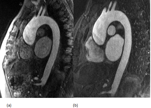
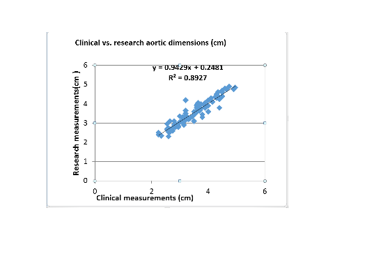
This study was IRB approved and informed consent was obtained. The study population consisted of 14 patients with known or suspected TAA presenting for routine follow up with CE MRA of the thoracic aorta. All patients underwent thoracic Gadobutrol enhanced MRA as a routine clinical scan and returned on average 5 weeks later for a research Gadoterate Meglumine MRA. The injection rate and volume were the same for both studies. Measurements of the aortic diameters were obtained at the levels of sinus of Valsalva, Sino-tubular junction, ascending aorta, proximal arch, distal arch and distal descending aorta for both scans. The average diameters for each measurement were calculated and correlation was assessed by linear regression analysis. Qualitative analysis consisted of two radiologists, blinded to the type of contrast used, scoring 4 arterial segments for image quality, signal-to-noise ratio (SNR) and contrast-to-noise ratio (CNR) using a 5-point Likert scale.Results
Qualitatively, there was no significant difference in image quality, SNR and CNR between both images for both reviewers (ICC>0.8). Quantitatively, there was an excellent agreement between aortic diameter measurements for both scans (r2=0.89, p<0.001) at all measured aortic levels.Discussion
Gadoterate Meglumine enhanced MRA showed comparable diagnostic image quality when analyzing the thoracic aorta in patients with thoracic aortic aneurysms with excellent correlation of aortic measurements when compared to Gadobutrol scans.Conclusion
Gadoterate Meglumine enhanced MRA is comparable to gadobutrol enhanced MRA for imaging patients with thoracic aortic aneurysms, providing adequate image quality and precise measurements of the aortic diameters.Acknowledgements
No acknowledgement found.References
1. Clouse WD, Hallett JW Jr, Schaff HV, Spitettell PC, Rowland CM, Illstrup DM, Melton LJ. Acute aortic dissection: population-based incidence compared with degenerative aortic aneurysm rupture. Mayo Clin Proc. 2004;79:176-180. 2. Laura A. Freeman, Phillip M. Young, Thomas A. Foley, Eric E. Williamson, Charles J. Bruce, Kevin L. Greason. CT and MRI Assessment of the Aortic Root and Ascending Aorta. American Journal of Roentgenology. 2013;200: W581-W592. 10.2214/AJR.12.9531 3. Kirchin MA, Runge VM. Contrast agents for magnetic resonance imaging: safety update. Top Magn Reson Imaging. 2003;14(5):426–35.Figures