3357
Comparison of Time-Resolved 3D Contrast-Enhanced MR Angiography on a Compact 3T Scanner with a Whole-Body 3T Scanner1Radiology, Mayo Clinic, Rochester, MN, United States
Synopsis
High spatiotemporal resolution contrast-enhanced MR angiography of the whole brain was performed on a compact 3T system with a 32 channel RF coil and compared to a spatial-resolution-matched study on a 60 cm bore whole-body 3T scanner. The quality of images from both scanners was excellent. Higher temporal resolution (4.18 s vs 5.75 s) on the compact 3T scanner was enabled by high performance gradients and increased PNS limits compared to the whole-body scanner.
Introduction
Since its advent in the early 1990s (1), contrast-enhanced MR angiography (CE-MRA) has benefitted from advances in acquisition and reconstruction to more accurately portray the temporal and spatial transit of contrast material through the vasculature. Specifically, advances in gradient hardware that allow for faster traversal of k-space and shorter scan times, parallel imaging techniques that facilitate reduced sampling, and advanced reconstruction schemes that leverage a priori knowledge of signal sparsity all combine to improve spatiotemporal resolution and signal-to-noise ratio (SNR). While virtually all modern MR scanners are compatible with parallel imaging and advanced reconstruction techniques, gradient hardware capabilities vary widely, with maximum gradient strength (Gmax) and gradient slew rate (SR) dependent on the gradient amplifier and the radius and length of the gradient coil. A compact 3T (C3T) MR system (2) with high performance gradients, allowing for reduced TE and TR, has recently been brought online at our institution. While shortening the TE and TR is advantageous from a temporal resolution standpoint, it is possible that the signal magnitude could suffer due to the reduced T1 recovery during the shorter TR. Further, like other modern scanners, this scanner is compatible with parallel imaging through the use of a 32 channel RF receive head coil. The purpose of this work is to perform high spatiotemporal resolution 3D CE-MRA of the brain using the C3T and compare – with spatial-resolution-matched acquisition – to exams performed on a whole-body, 60 cm bore 3T scanner.Methods
The C3T scanner used in this study features a lightweight and low-cryogen (12 liters) superconducting cylindrical-bore main magnet and high-performance gradient system (Gmax=80mT/m, SR=700 T/m/s). In addition to the high gradient performance, the smaller gradient size (42 cm inner diameter) allows for higher gradient switching rates without inducing peripheral nerve stimulation (PNS) (3). The linear and B0 concomitant fields of the asymmetric gradient design were corrected by real-time gradient pre-emphasis (4) and frequency tracking (5), respectively. In this IRB-approved study, healthy volunteers were imaged with the 32-channel head coil (Nova Medical, Wilmington, MA) on both the compact 3T scanner and a whole-body 3T scanner (MR750, GE Healthcare, Waukesha, WI) capable of 50 mT/m and 200 T/m/s. Typical scan parameters are shown in Table 1, but did vary to accommodate different volunteer sizes. Protocols between the two scanners were matched in spatial resolution (approximately 1mm isotropic), but were allowed to vary in temporal resolution (due to different gradient capabilities). Images were reconstructed with an iterative sparse reconstruction (6,7) and image quality was compared qualitatively for this initial report.Results
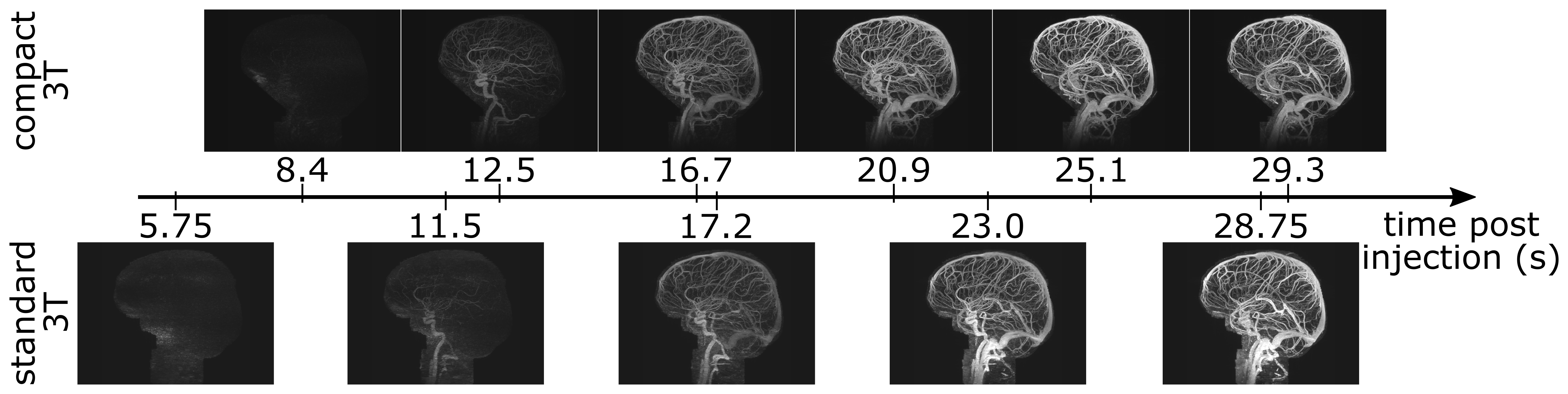
The studies were performed successfully, and no PNS was reported by any volunteer. Sagittal maximum-intensity-projection images (MIPs) of time-resolved studies from the compact 3T scanner (a) and whole-body 3T scanner (b) are shown in Figure 1. The quality of the images from both exams is excellent; note that with the same spatial resolution, the images acquired on the C3T scanner have higher temporal resolution and were able to capture an arterial phase before venous enhancement.
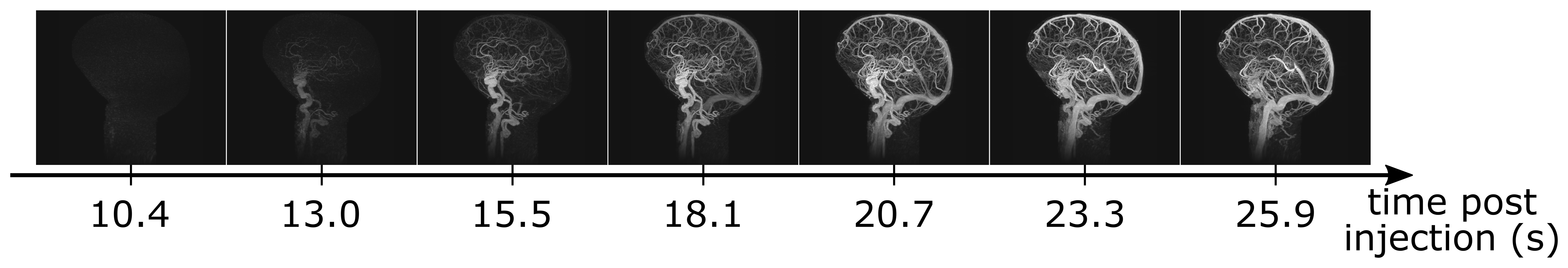
A time series of sagittal MIPs is shown in Figure 2. An image update time of 2.6 s was achieved on the C3T, while on the whole-body scanner with the same parameters the update time was 3.7 s. This volunteer has not yet returned for imaging on the whole-body scanner.
Discussion
An approximately 31% reduction in TE and TR, and therefore image update time, compared to a 60 cm bore whole-body 3T system was observed when using the C3T system (Table 1), allowing for high spatial and temporal resolution in the 3D time-resolved exam. Future work includes comparative assessment of additional subjects on both scanners with specific evaluation of image quality, vessel conspicuity, spatial resolution, and temporal fidelity between images from the two scanners.Acknowledgements
The authors would like to acknowledge study coordinator Kathy Brown and the following sources of funding: NIH EB000212, NIH RR018898, NIH BRP-R01-EB010065, NIH U01-EB024450.References
- Prince MR et al. J. Magn. Reson. Imaging 1993;3:877–881.
- Foo T. In: Proceedings of the 24th Annual Meeting of ISMRM. Singapore; 2016. p. 3629.
- Chronik BA et al. Magn. Reson. Med. 2001;46:386–394.
- Tao S et al. Magn. Reson. Med. 2017;77:2250–2262.
- Weavers PT et al. Magn. Reson. Med. Early View. DOI: 10.1002/mrm.26790.
- Trzasko JD et al. Magn. Reson. Med. 2011;66:1019–1032.
- Trzasko JD et al. In: Proceedings of the 23rd Annual Meeting of ISMRM. Toronto, Canada; 2015. p. 574.
Figures