3350
Combined Assessment of Peripheral Artery Disease by MRI-based Vascular Calcification Visualization and Quiescent Interval Single-Shot (QISS) MRA1Department of Radiology, Medical University of South Carolina, Charleston, SC, United States, 2Department of Medicine, Medical University of South Carolina, Charleston, SC, United States, 3NorthShore University, Evanston, IL, United States
Synopsis
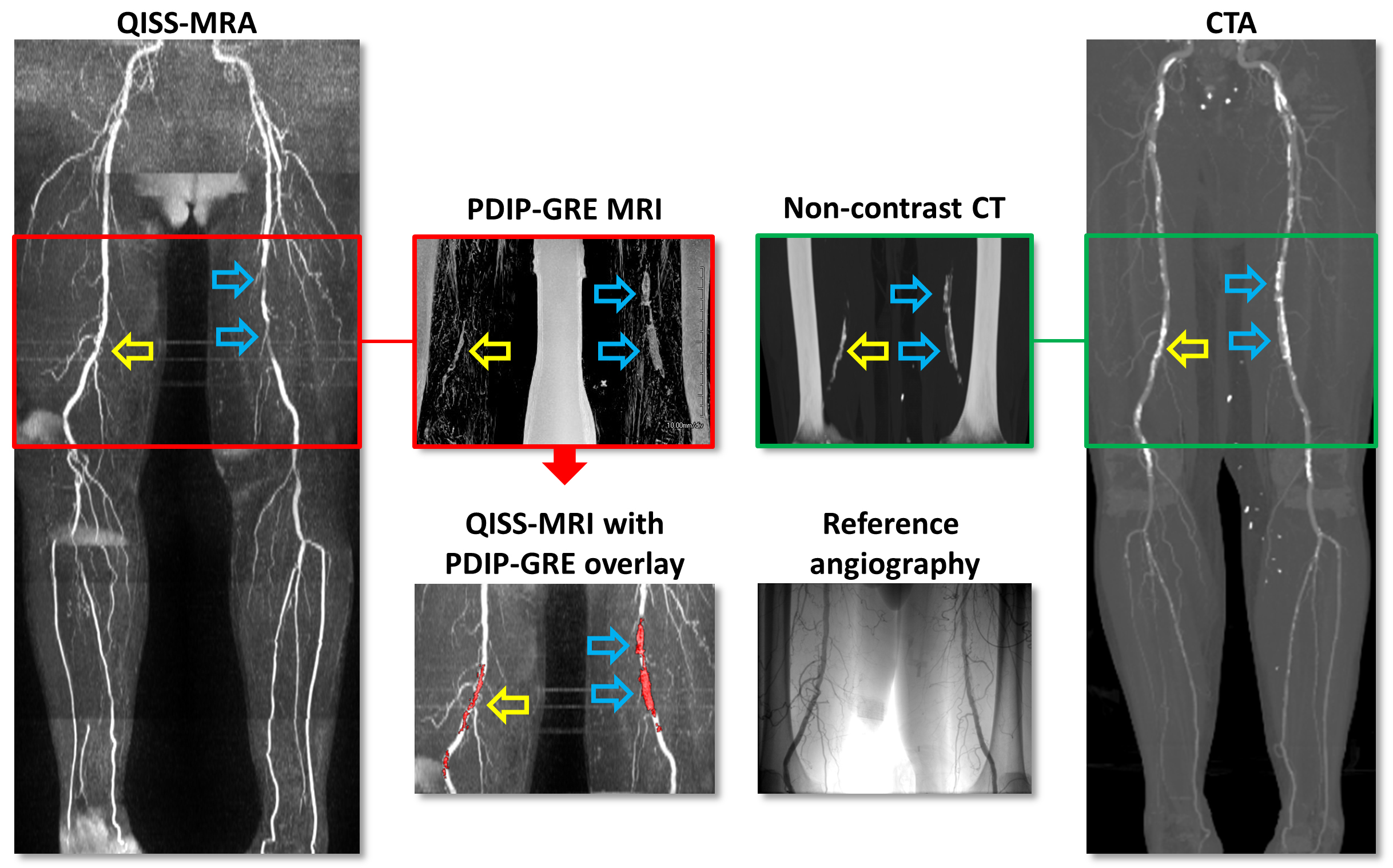
The diagnostic accuracy of quiescent-interval single-shot (QISS) MRA to detect peripheral artery disease (PAD) has been shown to be similar to that of CTA. Unlike CTA, standard MR techniques are limited in the detection of vascular calcification. However, proton density-weighted, in-phase 3D stack-of-stars gradient-echo (PDIP-GRE) prototype pulse sequence has been shown to accurately depict calcifications in PAD. In our study, PDIP-GRE MRI provided comparable assessment to CTA. The MRI visualization of lower extremity vascular calcification improved readers’ confidence and the diagnostic accuracy of QISS-MRA in detecting significant vascular stenoses. Quantification of vascular calcium with MRI showed good agreement with CTA.
Introduction
The contrast administration-related risk and other technological advances have increased the interest in noncontrast MRA techniques, especially in patients with peripheral artery disease (PAD). A recent technique, quiescent interval single-shot (QISS) MRA, has been introduced for the imaging of the lower extremity arterial system and shows high diagnostic accuracy for the detection of significant vascular stenosis in patients with PAD.1,2 However, QISS MRA is unable to visualize vascular calcification. A novel proton density-weighted, in-phase 3D stack-of-stars gradient-echo (PDIP-GRE) prototype pulse sequence, however, seems to be promising to overcome of this limitation.3 In this study we aimed to investigate the feasibility of a combined QISS MRA and PDIP-GRE MR imaging (MRI) protocol for the visualization of vascular stenosis and calcification in patients with PAD, using CTA and digital subtraction angiography (DSA) as the reference.Methods
Fourteen PAD patients (68±9 years) referred for lower extremity computed tomography angiography (CTA, Siemens Force; FOV 350 mm; pitch 0.7; collimation, 2 × 64 × 0.6 mm for both detectors; and tube voltage and current, 150 kV/59 reference mAs for tube A and 90 kV/95 reference mAs for tube B) prior to digital subtraction angiography (DSA) (Siemens Axiom Artis), were prospectively enrolled for a same-day 1.5T MRI (Siemens Avanto). Prototype QISS-MRA (FOV 400×260mm2, TR/TE 3.5/1.4ms, flip angle 90°, in-plane resolution 1.0×1.0mm2) was performed covering the abdominal aorta and lower extremity run-off. Prototype PDIP-GRE MRI (FOV 416mm, TR/TE 9.6/4.7ms, flip angle 4.5°, radial views 660) was acquired covering the iliofemoral region. Image evaluation was performed on a per-segment basis according to an 18-segment model. Two readers rated diagnostic confidence (3-point scale) and graded stenosis (< or >50%) on QISS-MRA without and with the visualization of calcification. Sensitivity and specificity were calculated using the McNemar-test with DSA as reference. Intra-arterial calcium was quantified using ImageJ (NIH) and compared between MRA and CTA using paired t-test and Bland-Altman analysis.Results
A total of 252 vascular segments were evaluated with QISS-MRA and 84 segments were available for calcification assessment. Of those 84 segments, >50% stenosis was detected in 37 (44%), while calcium was present in 58 (69%). The diagnostic confidence (2.1 [1.9-2.3] vs. 2.3 [2.1-2.5]; P<0.0001) and the sensitivity and specificity of QISS-MRA in the detection of >50% stenosis (83.2% and 97.6% vs. 86.4% and 98.0%, respectively) were improved when calcification data were provided to the readers. Quantification of calcification showed no statistical difference between MRI and CTA (121±77mm3 vs 127±84mm3; P=0.0249), and Bland-Altman analysis revealed excellent agreement with a mean of differences at -5.8mm3.Discussion
Both CTA and MRA techniques can be used when outlining revascularization strategies. The advantage of CTA over currently available MRA techniques is the ability of visualizing intra-arterial calcifications, although, blooming artifacts in calcified areas may influence the assessment of stenosis significance. The PDIP-GRE MRI technology, however, provides comparable assessment to CTA. The MRI visualization of lower extremity vascular calcification improved readers’ confidence and the diagnostic accuracy of QISS-MRA in detecting significant vascular stenoses. Quantification of vascular calcium with MRI showed good agreement with CTA.Conclusion
Based on our initial results, the PDIP-GRE MRI technique seems to be promising for the complex assessment of vascular anatomy in PAD, which could be beneficial in interventional procedure planning.Acknowledgements
References
1. Edelman RR, Sheehan JJ, Dunkle E, Schindler N, Carr J, Koktzoglou I. Quiescent-interval single-shot unenhanced magnetic resonance angiography of peripheral vascular disease: Technical considerations and clinical feasibility. Magn Reson Med 2010;63:951-8.
2. Varga-Szemes A, Wichmann JL, Schoepf UJ et al. Accuracy of Noncontrast Quiescent-Interval Single-Shot Lower Extremity MR Angiography Versus CT Angiography for Diagnosis of Peripheral Artery Disease: Comparison With Digital Subtraction Angiography. JACC Cardiovasc Imaging 2017;10:1116-1124.
3. Ferreira Botelho MP, Koktzoglou I, Collins JD et al. MR imaging of iliofemoral peripheral vascular calcifications using proton density-weighted, in-phase three-dimensional stack-of-stars gradient echo. Magn Reson Med 2017;77:2146-2152.
Figures