3341
Whole Body Vascular MR Imaging in Five Minutes for Patients With ClaustrophobiaPuja Shahrouki1,2, John Moriarty1,2, Biraj Bista1,2, Sarah Khan1,2, Stephen Kee1, Brian DeRubertis3, Takegawa Yoshida1,2, Kim-Lien Nguyen2,4, and J. Paul Finn1,2
1Department of Radiological Sciences, David Geffen School of Medicine at UCLA, Los Angeles, CA, United States, 2Diagnostic Cardiovascular Imaging Laboratory, David Geffen School of Medicine at UCLA, Los Angeles, CA, United States, 3Department of Cardiothoracic Surgery, David Geffen School of Medicine at UCLA, Los Angeles, CA, United States, 4Division of Cardiology, David Geffen School of Medicine at UCLA and VA Greater Los Angeles Healthcare System, Los Angeles, CA, United States
Synopsis
Patients with claustrophobia represent a significant proportion of patients who would otherwise be suitable candidates for MR angiography. With conventional acquisition techniques, examination times for MRA typically exceed 30 minutes and claustrophobic patients are often unwilling or unable to undergo the study. We implemented a new approach to minimize time in the magnet bore for patients with claustrophobia, acquiring comprehensive vascular evaluation of the thorax, abdomen and pelvis in as little as 5 minutes.
Introduction
Whereas MR angiography has been widely used for more than two decades, the rapid proliferation of CT angiography has underscored the advantages in speed and practicality of modern CTA when compared to contrast enhanced MR angiography (CEMRA). Beyond challenges in workflow, the relatively long magnet time associated with conventional MR techniques serves to discourage patients with even modest levels of claustrophobia. Furthermore, recent controversies surrounding the safety of gadolinium based contrast agents (GBCA) has added further uncertainty in the cost-benefit balance point of conventional CEMRA. We hypothesize that with the use of ferumoxytol (Feraheme, AMAG Pharmaceuticals), comprehensive vascular imaging of the thorax, abdomen and pelvis can be performed in as little as 5 minutes within the magnet bore, sufficient to reassure all but the most claustrophobic patients and greatly improving workflow and throughput.Methods
In this HIPAA-compliant and IRB-approved study, all patients expressed reluctance to undergo the MR examination due to claustrophobia, but agreed to a trial of up to 10 minutes in the scanner bore. Following localizer sequences, breath held, high-resolution 3-D ferumoxytol enhanced MR angiography (FEMRA) was carried out in one (n=1) or two (n=12) overlapping stations on a 3.0T MR system (Magnetom TIM Trio or Magnetom Skyra, Siemens Medical Solutions) or a 1.5T MRI system (Magnetom TIM Avanto, Siemens Medical Solutions). Acquisition time for each station was 16-20 seconds and voxel dimensions were on the order of 1 mm x 1.2 mm x 1.3 mm. Overlapping stations were composed into one large field of view study using vendor software (Image Compose, Siemens). The total scanning time was measured from the beginning of localizer image acquisition to the end of the ultimate FEMRA acquisition. Scanner tuning and coil adjustment routines preceded localizer acquisition. Patient-specific machine shimming was not performed and the default shim settings were employed to minimize adjustment time.Results
Thirteen examinations were carried out in 12 claustrophobic patients (age 11 to 84 years, 6 females) with renal failure (n=11) or hemorrhagic hereditary telangiectasia (n=1), who underwent IV infusion of 4 mg /kg of ferumoxytol (AMAG Pharmaceuticals, Waltham, MA) outside the magnet bore. All examinations were completed successfully without adverse events. The imaging indications included central venous occlusion (n=8), aortic stenosis (n=2), central arterial occlusion (n=1), or hereditary hemorrhagic telangiectasia (n=1). The average scanning time was 6.44 minutes (range 4.16 to 10.10 minutes) including tune up time which was less than one minute. All scans were considered of high diagnostic quality and supported full visualization of arterial and venous anatomy from the neck to the thighs.Conclusion
Comprehensive vascular imaging of the thorax, abdomen and pelvis can be completed in as little as 5 minutes in the scanner bore using focused MR angiographic image acquisition, following infusion of ferumoxytol. The implications for imaging of claustrophobic patients and for patient throughput are profound.Acknowledgements
No acknowledgement found.References
1. Dewey M, Schink T,Dewey CF. Claustrophobia during magnetic resonance imaging: cohort study in over 55,000 patients. J Magn Reson Imaging. 2007;26:1322-7Figures
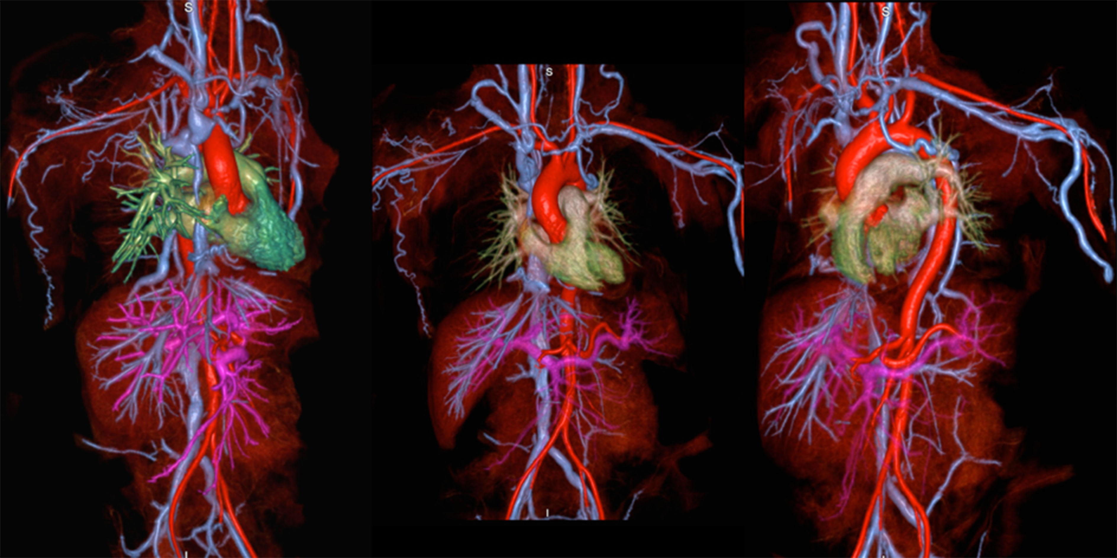
Figure 1.
Reconstructed 3D FE-MRV volume rendered images summarize
vascular anatomy from the neck to the thighs.
Source images were acquired in two breath holds. This patient has occlusive disease of the
right upper extremity and central thoracic veins (rendered in blue).