3340
MR Venography with Ferumoxytol in Central Venous Occlusion1Department of Radiological Sciences, David Geffen School of Medicine at UCLA, Los Angeles, CA, United States, 2Diagnostic Cardiovascular Imaging Laboratory, David Geffen School of Medicine at UCLA, Los Angeles, CA, United States, 3Department of Cardiothoracic Surgery, David Geffen School of Medicine at UCLA, Los Angeles, CA, United States, 4Division of Cardiology, David Geffen School of Medicine at UCLA and VA Greater Los Angeles Healthcare System, Los Angeles, CA, United States
Synopsis
Treatment of central venous occlusion is guided largely by anatomic considerations determined by pre-procedural imaging. Current approaches to imaging such as ultrasound and conventional cross-sectional imaging of central veins face many technical challenges and may be contraindicated in patients with renal impairment. We demonstrated that ferumoxytol-enhanced MR venography (FE-MRV) is a safe and highly accurate diagnostic tool that can be used as a reliable pre-interventional vascular map.
Introduction
As therapy prolongs survival in patients with cancer and organ failure, central venous obstruction is an increasingly common and potentially life threatening complication of venous instrumentation. Treatment of venous occlusion is guided largely by anatomic considerations and imaging of central veins is challenging. Ultrasound is of little value in the chest and conventional CT and MRI often struggle with suboptimal venous enhancement from extracellular contrast agents. Conventional iodinated and gadolinium-based contrast agents may be less desirable in patients with renal failure. We aim to evaluate the diagnostic performance of FE-MRV in central venous occlusion and to assess its clinical impact on patient management.Methods
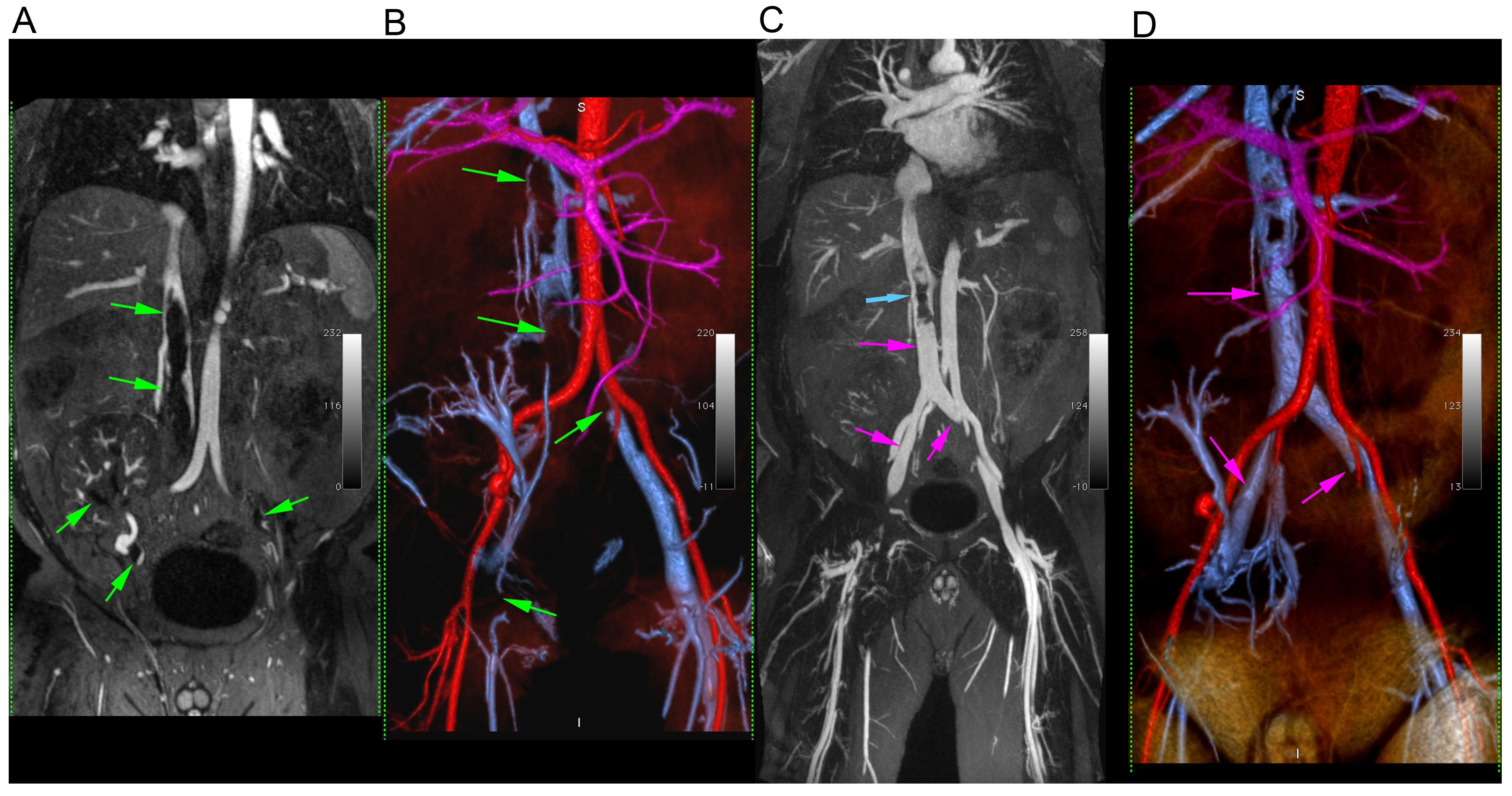
Fifty-two consecutive adult patients (median age 47, interquartile range (IQR) 32 – 61 years; 29 male) with suspected venous occlusion underwent FE-MRV on a 3.0T MRI system (Magnetom TIM Trio, Magnetom Prisma Fit or Magnetom Skyra, Siemens Medical Solutions) or 1.5T MRI system (Magnetom TIM Avanto, Siemens Medical Solutions) between June 2013 and May 2017. Indications included: chronic kidney disease (n=44), acute kidney injury (n=4) or anticipated failure of alternative techniques (n=4). All patients underwent infusion of ferumoxytol, 4 mg /kg, with continuous monitoring of heart rate, blood pressure and pulse oximetry throughout the examination. Breath-held, high resolution steady state FE-MR angiography was acquired through the chest, abdomen and pelvis, with parameters adjusted to patient size and ability to cooperate. Typical acquisition times per station were 16-22 seconds and voxel dimensions were typically 1 x 1.2 x 1.4 mm. Two blinded reviewers independently scored FE-MRV exams for twenty-one named venous segments on a 4-point scale, where scores ≥2 were considered of diagnostic quality. All segments of diagnostic quality within the field of view were analyzed for occlusion. Disagreements were resolved by consensus of the primary reviewers with a third senior reviewer. Correlative conventional venography in a subset of 15 patients was used as the reference standard for determination of diagnostic accuracy. Retrospective chart review was conducted to determine clinical impact of FE-MRV with regards to need for additional imaging, outcome of interventional procedures and renal function. Differences between pre and post MRI creatinine were determined by using two-tailed t-test. Interobserver agreement was determined by using Gwet’s AC1 statistic, with the following grading: 0.00-0.20, poor; 0.21-0.40, fair; 0.41-0.60, moderate; 0.61-0.80, good; 0.81-1.00, very good.Results
All patients underwent FE-MRV without any adverse events. 99.5% (1033/1038) segments were considered of diagnostic quality by both observers with very good interobserver agreement (AC1=0.91). The interobserver agreement for venous occlusion was also very good (AC1=0.93). Only 5% of segments had to be resolved by consensus. The overall sensitivity, specificity, positive predictive and negative predictive value was 0.793, 0.944, 0.852 and 0.918 respectively. There was no significant difference in the pre-MRI and post-MRI creatinine (p>0.05). Only two patients (8 %) had additional imaging after the FE-MRV prior to an intervention based on the clinician’s request. In both cases, additional imaging confirmed the FE-MRV findings. Twenty-five successful FE-MRV-guided endovascular interventions were carried out in 24 patients without complications.Conclusion
FE-MRV is a practical and highly accurate technique for mapping of central thoracic and abdominal veins and can be used to inform image guided therapy. As a technique for venous imaging, FE-MRV may play a pivotal role in the care of patients in whom conventional contrast agents are contraindicated or ineffective.Acknowledgements
No acknowledgement found.References
1. Modabber M, Kundu S. Central venous disease in hemodialysis patients: an update. Cardiovasc Intervent Radiol 2013;36(4):898-903.
2. Finn JP, Nguyen KL, Han F, et al. Cardiovascular MRI with ferumoxytol. Clin Radiol 2016;71(8):796-806.
3. Gwet KL. Computing inter-rater reliability and its variance in the presence of high agreement. Br J Math Stat Psychol. 2008;61:29-48.
Figures