3336
The assessment of global and regional strain in patients with preserved ejection fraction after Fontan operation using feature tracking technique as compared with healthy children1Radiology, Shanghai Children's Medical Center, Shanghai, China
Synopsis
The quantification of myocardial deformation may allow detection of early abnormalities and provide independent prognostic information, as demonstrated in echocardiographic studies.Meanwhile, some studies have suggested that CMR-FT may be evaluated earlier than ejection fraction to detect early abnormalities of the ventricular myocardium in postoperative follow-up of CHD. But, there is still limited experience with cardiac magnetic resonance feature tracking strain analysis in child patients. To the best of our knowledge, the significance of quantifying ventricular myocardial deformation in post-Fontan patients with pEF using CMR-FT has not been investigated. Therefore, the aim of this study was to evaluate the myocardial strain in children with pEF after the Fontan operation using feature tracking technique compared to healthy children.
Purpose:
The purpose of the study was to evaluate the myocardial strain in children with preserved ejection fraction(pEF) after the Fontan surgery using feature tracking(FT) technique.Methods:
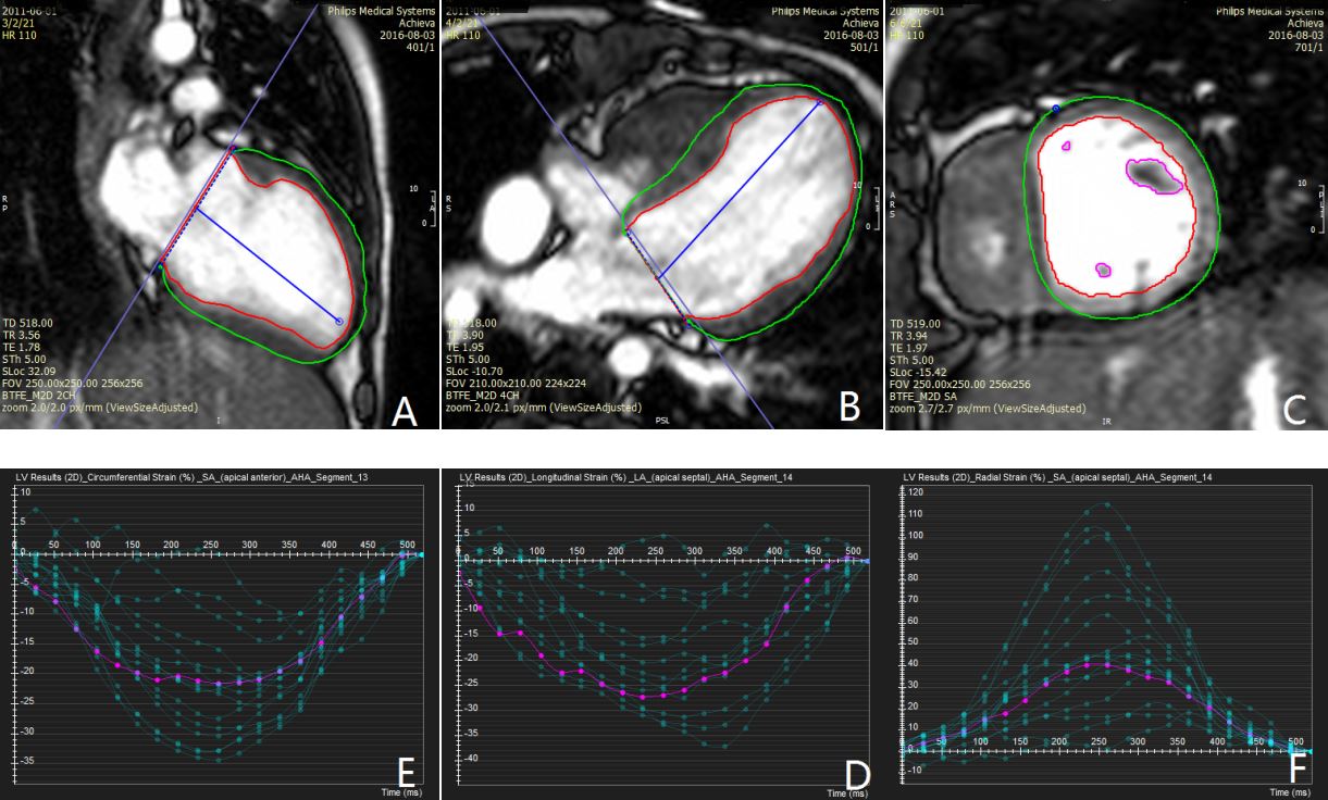
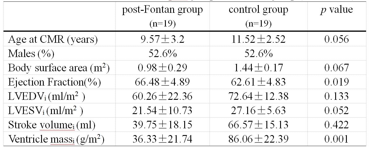
Clinical cardiac magnetic resonance (CMR) studies between 1/2015 and 8/2017 in 19(male/female,10/9) patients with Fontan circulation and 19 age- and gender-matched healthy children volunteers were retrospectively enrolled for the study. CMR imaging was conducted on a 1.5 Tesla MRI scanner. Conventional ventricular function parameters are calculated in two groups. Meanwhile, global and regional strain of the left ventricle in post-Fontan group(pEF) and the left ventricle in the control group were obtained using a CMR-FT software. The comparison between two groups was assessed by the Mann-Whitney test. The correlation between ejection fraction and ventricular strain was performed using Pearson’s correlation. Bland-Altman method identified coefficients of variation for the global strain between two observers on GraphPad prism 5 software.Results:
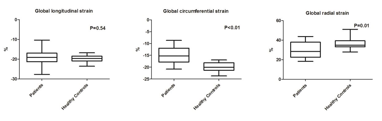
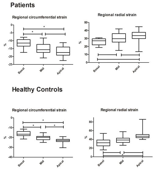
To compare post-Fontan and control groups, LV global longitudinal strain (GLS) was reduced to -18.87±4.61 in post-Fontan group (-19.72±1.58 in controls, p = 0.54), and global circumferential strain (GCS) reduced to -14.55±3.79 (-19.91±1.97 in controls, p < 0.001). The global radial strain (GRS) was also reduced at 29.62±8.41 (36.85±5.95 in controls, p = 0.01). The regional circumferential strain decrease was marked in the basal segments in the post-Fontan group (mid, p = 0.005, apical, p < 0.001). The global circumferential strain correlated well with LVEF (r =−0.49, p =0.03). The global strain intra- and inter-observer yielded a good agreement except for global radial strain in the post-Fontan group.Discussion and Conclusion:
The assessment of cardiac strains on standard cine CMR image is feasible in patients with Fontan operation. The patients with pEF showed reduced cardiac strains compare to control groups. The global circumferential strain can early detect abnormal myocardial function with pEF in the post-Fontan group. Further studies are needed to assess its value for diagnosis using feature tracking after Fontan operation, especially in combination with ventricular twisting and dyssynchrony.Acknowledgements
The authors appreciate Hai-tao You and Tong-tong Han, Circle Imaging Systems, Circle CVI Corporation Canada for technical assistance.References
1. Pedrizzetti G, Claus P, Kilner P J, et al. Journal of Cardiovascular Magnetic Resonance, 2016, 18(1): 51.
2. Augustine D, Lewandowski A J, Lazdam M, et al. Journal of Cardiovascular Magnetic Resonance, 2013, 15(1): 8.
3. Haeck M L A, Scherptong R W C, Marsan N A, et al. Circulation: Cardiovascular Imaging, 2012, 5(5): 628-636.
Figures