3321
Whole heart First-pass spiral perfusion imaging with 1.25mm resolution at 3T1Medicine, University of Virginia, Charlottesville, VA, United States, 2Radiology, University of Virginia, Charlottesville, VA, United States, 3Biomedical Engineering, University of Virginia, Charlottesville, VA, United States
Synopsis
First-pass contrast-enhanced myocardial perfusion imaging is a useful noninvasive tool to evaluate patients with coronary artery disease, but current techniques are still limited in spatial-temporal resolution, and ventricular coverage which reduces the sensitivity to detect perfusion differences between the endocardium and epicardium and quantify ischemic burden. Outer-volume suppression (OVS) can achieve good signal suppression around the heart, but may have SAR limitations at 3T. In this study, we designed a spiral pulse sequence with slice-interleaved or simultaneous multi-slice (SMS) acquisition without OVS to achieve comparable high quality ultra-high 1.25mm resolution perfusion imaging. The sequences were tested in heathy volunteers and demonstrated high image quality.
Introduction
First-pass contrast-enhanced myocardial perfusion imaging is a useful noninvasive tool to evaluate patients with coronary artery disease (CAD)1. However, current first-pass perfusion imaging techniques still have limited spatial resolution (~2 mm) which may reduce the sensitivity to detect perfusion differences between the endocardium and epicardium. We recently demonstrated perfusion imaging with an ultra-high spatial resolution of 1.25 mm by using outer-volume suppression (OVS)2, simultaneous multi-slice (SMS) imaging3, and dual density spiral trajectories at 3T4. However, the OVS module requires a high B1 field to achieve good suppression performance which will increase SAR and limit the application of the technique for adenosine stress perfusion imaging. Thus, the goal of this study is to further develop these spiral pulse sequences to achieve ultra-high spatial resolution perfusion imaging at 3T without OVS.
Methods
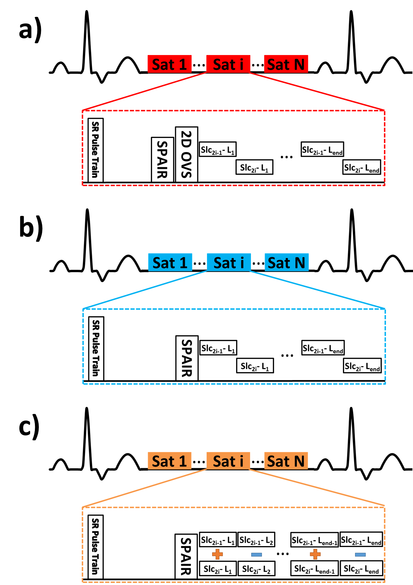
Three different pulse sequences were evaluated in this study, including an interleaved acquisition of two slices per saturation recovery (SR) block with OVS (Fig 1a) and without OVS (Fig 1b), and SMS with a multi band factor of 2 per SR without OVS (Fig 1c). To further reduce the SAR, an optimized 5 hard-pulse SR was utilized5. Detailed parameters of three sequences are listed in Table 1. Resting first-pass perfusion using the proposed sequences were performed with a 0.075 mmol/kg Dotarem bolus, separated by 20 min contrast washout time, in 5 healthy subjects on a 3T Prisma Siemens scanner. One subject was imaged using interleaved acquisition with and without OVS, while all other subjects were imaged by interleaved acquisition without OVS and SMS without OVS. The images were reconstructed by L1-SPIRiT or SMS-L1-SPIRiT using finite temporal difference as the sparsity transform6. The image reconstruction was formulated as the following optimization problem: $$ argmin_x \| \Phi DFx - y \|^2 + \lambda_1 \|(G-I)x\|^2 + \lambda_2\|\Psi x\|_1 $$ Where $$$F$$$ transfers the data from image domain to k-space domain, $$$D$$$ is the inverse gridding operator that transfers the Cartesian grid to spiral trajectory, $$$G$$$ is an image-space SPIRiT operator that represents the k-space self-consistency convolutions in the image domain, $$$\Psi$$$ is the finite time difference transform that operates on each individual coil separately to achieve sparsity in the temporal domain of image time series, $$$\Phi$$$ is the operator to combine multiple phase modulation slices to single SMS slice for SMS acquisition or $$$\Phi=I$$$ or interleaved acquisition, $$$\lambda_1$$$ and $$$\lambda_2$$$ are parameters that balance the data acquisition consistency with calibration consistency and sparsity. An upfront scan without SMS is used to derive the calibration kernel. The overall image quality was graded by a single cardiologist based on 5 point scale (5-excellent, 1-poor).Results
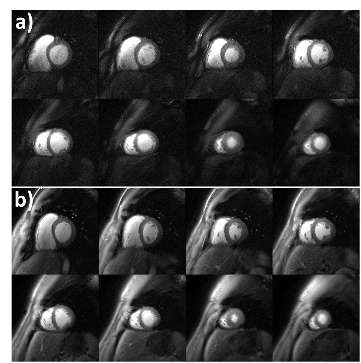
Figure 2 shows the direct comparison of whole heart perfusion imaging at a middle time frame during first pass using interleaved acquisition with OVS (a) and without OVS (b) from the same healthy subject. The OVS can achieve good signal suppression outside the heart but requires higher SAR to achieve the necessary B1 field. Good image quality is achieved at 1.25mm spatial resolution with or without OVS. Reduction of the acquisition time per interleaf from 5ms to 4ms improved image quality and reduced drop-out artifacts as compared to our prior study. Figure 3 presents another example case with the same resolution at a similar time frame during first-pass of contrast using an interleaved acquisition pulse sequence without OVS. Figure 4 shows perfusion images from the same subject acquired using the SMS MB=2 pulse sequence without OVS. Image quality scores were 4.6±0.2 and 4.0±0.7 for the interleaved and SMS techniques without OVS respectively. Either an interleaved acquisition or SMS strategy can produce high quality perfusion images with whole heart coverage and ultra-high spatial resolution without requiring the OVS module, which may improve applicability for stress perfusion imaging.Conclusion
We demonstrated the successful application of ultra-high resolution spiral perfusion sequence using interleaved acquisition and SMS techniques either with or without OVS. High-resolution whole-heart perfusion will potentially serve as a tool to quantify regional differences in perfusion of the subendo and subepi myocardium. Further validation will be required in patients undergoing adenosine stress CMR.Acknowledgements
This work was supported by NIH K23 HL112910 and R01 HL079110.References
1. Nagel E, Klein C, Paetsch I, Hettwer S, Schnackenburg B, Wegscheider K, Fleck E. 2003. Magnetic resonance perfusion measurements for the noninvasive detection of coronary artery disease. Circulation. 108(4):432-437.
2. Yang Y, Zhao L, Chen X, Shaw PW, Gonzalez JA, Epstein FH, Meyer CH, Kramer CM, Salerno M. 2017. Reduced field of view single-shot spiral perfusion imaging. Magnet Reson Med.
3. Barth M, Breuer F, Koopmans PJ, Norris DG, Poser BA. 2016. Simultaneous multislice (sms) imaging techniques. Magn Reson Med. 75(1):63-81.
4. Yang Y, Van Houten M, Kramer CM, Salerno M. 2017. Ultra-high spatial resolution spiral myocardial perfusion imaging with whole heart coverage at 3T. Accept for 2018 SCMR/ISMRM workshop.
5. Chow K, Kellman P, Spottiswoode BS, Nielles-Vallespin S, Arai AE, Salerno M, Thompson RB. 2015. Saturation pulse design for quantitative myocardial t1 mapping. Journal of Cardiovascular Magnetic Resonance 17:84.
6. Yang Y, Van Houten M, Norton P, Hagspiel K, Kramer CM, Mugler JP, Salerno M. Spiral Simultaneous Multi-Slice First-Pass Myocardial Perfusion Imaging. Proc Intl Soc Mag Reson Med 2017;25:3242
Figures