3317
Evaluation of geometric models for estimating left ventricular (LV) mass in 980 children using cardiac cine magnetic resonance imaging1Diagnostic and Interventional Radiology, Baylor St Luke's Medical Center, Houston, TX, United States, 2Texas heart institute, Houston, TX, United States
Synopsis
LV mass computed from commonly used bi-plane and tri-plane ellipsoidal models can deviate significantly when compared to LV mass estimated from a stack of short axis balanced SSFP cine MR images. The results from the study show that by using different geometric assumptions for the shape of the endocardium (Cut-cone+cone) and epicardium (Cut-cone+parabola), it is possible to estimate LV mass with just two projections that is comparable to that obtained from a full stack of short axis slices.
INTRODUCTION
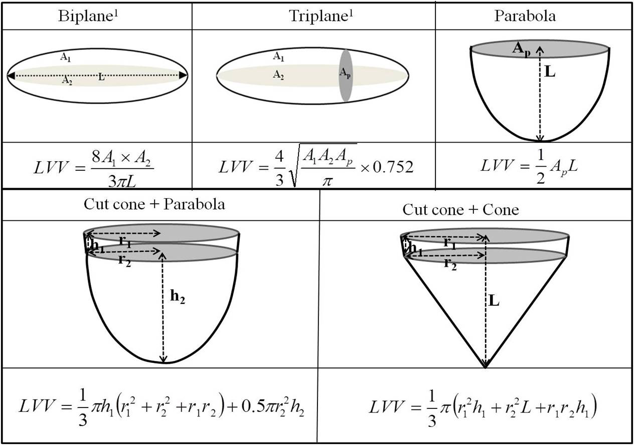
LV mass calculated from the difference between epicardial and endocardial LV volumes is an independent predictor of risk in cardiovascular disease. Echocardiography and x-ray angiographic methods estimate LV volume from few select projections by making geometric assumptions about the shape of LV. In contrast cardiac MRI (CMR) LV mass is calculated by delineating endo- and epicardial contours on a stack of cine slices in the short-axis (SAx) orientation without making any geometric assumptions. Studies have shown that these CMR measurements are both accurate and precise1,2. However, acquiring the SAx stack is time consuming and challenging for patients with compromised respiratory function. Previously we considered three new geometric models (in addition to the conventional biplane and triplane models) to capture the shape of LV (Figure 1) and demonstrated that LV volumes estimated from a sub-set of projections were comparable to the values obtained from a fully sampled dataset3. However these models were not evaluated for the estimation of LV mass.
In this work, we show that by modeling the endocardium and epicardium of the LV with distinct shapes, it is possible to obtain a reliable estimate of LV mass from as few as three projections and comparable to conventional geometric bi-plane and tri-plane models of the LV.
METHODS
Patient population: Data acquired from 980 middle and high school kids (recruited as a part of an IRB approved sudden cardiac death screening study) with no known history of heart disease (578 male, age: 12.6±1.1yrs, range: 10-15years) were used in this analysis.
MRI acquisition: VCG gated cine balanced steady state free precession images of the LV (spatial resolution 2x2x8mm3; temporal resolution < 50 ms) were acquired within SAx covering the whole LV, left ventricular outflow tract (LVOT) and horizontal four chamber (4CH) orientations using a 32 Channel RF coil for signal reception.
Data analysis: Endocardial and epicardial borders were circumscribed on the stack of SAx slices acquired at end-diastole (ED). Furthermore, the cross sectional endo- and epicardial areas of the SAx slice which included the dominant papillary muscle (Ap) was also recorded. The length of the left ventricle (measured from mitral annulus valve plane to the endocardium of the apex) from 4CH and LVOT orientations, as well as the diameter of the left ventricle for both epi- and endo- myocardium at the level of the mitral annulus in the 4CH and LVOT views was also recorded.
LV models: In addition to the widely used biplane and triplane models of the LV, we propose three additional models (parabola, cutcone+prabola and cutcone+cone) to account for the shape of the LV. From this basis set, a total of fifteen models were constructed by considering independent models for the endo- and epicardium (Figure 2).
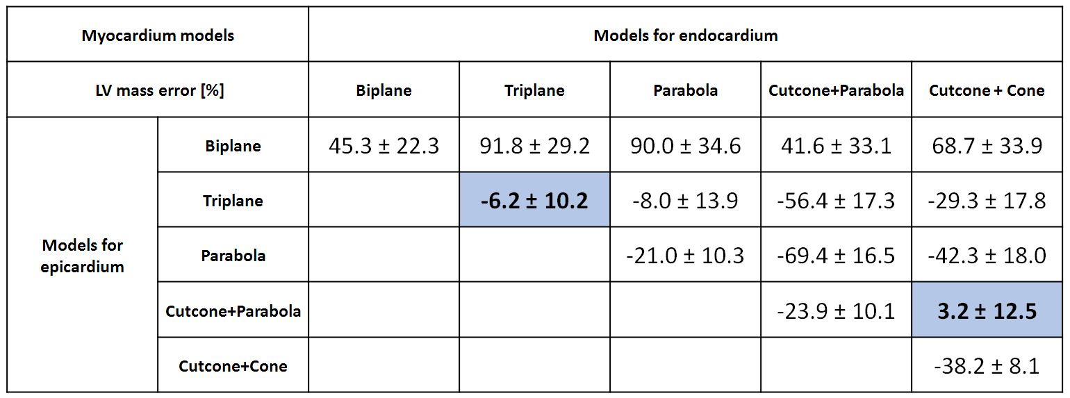
Data analysis: LV mass estimated from the stack of SAx slices was considered as the reference measurement. Five models used the same geometric model for both epicardium and endocardium volume to calculate the LV mass and the remaining ten models used a different geometric model for epicardium and endocardium. Bland-Altman analysis of the percentage-error in the calculation of LV mass compared to the reference measurement of all fifteen models is shown in Figure 2. (Expressed as bias ± 1 standard deviation ).
RESULTS
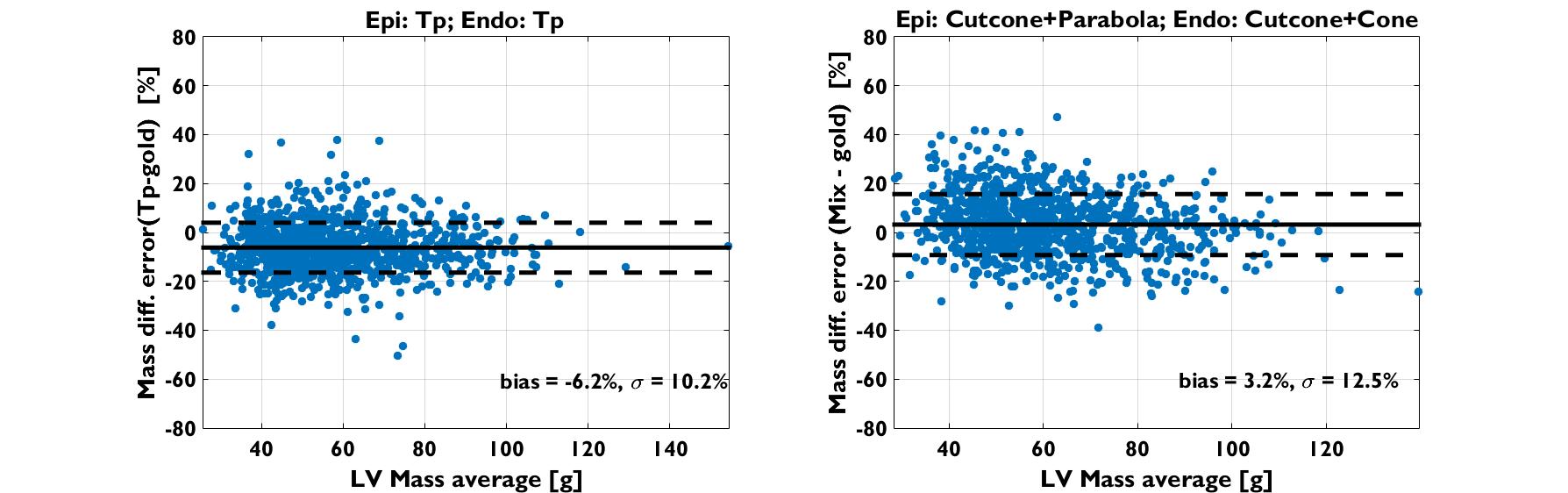
The average LV mass for this subject cohort calculated from fully sampled SAx data set is 60.7±17.9g. The main findings of this study are: Based on the result from Figure 2 and Bland-Altman plot of all data point for LV mass estimated from Triplane model and one best performed mixing model (Figure 3),
1. Among the mixed models (different model for end- and epicardium), the Cutcone+Parabola model for epi- and Cutcone+cone model for endocardial volumes yielded the least error for estimating LV mass (3%).
2. Bland-Altman analysis show that conventional triplane model underestimates LV mass by 6.2%, and the mixed model (endo: Cutcone+Cone, Epi: Cutcone+Parabola) overestimates LV mass by 3.2%.
3. Conventional geometric models: Biplane and Triplane model of the LV overestimate LV mass by 45% and underestimates LV mass by 6% respectively.
4. When the shape of the LV was assumed to be same for both endo-and epicardium, the LV mass calculated from the three proposed models resulted in substantial underestimation of LV mass (Figure 2).
DISCUSSION AND CONCLUSIONS
1. In this cohort of young children, our results show that the estimation of LV mass using conventional geometric models such as biplane and triplane can result in significant errors.
2. Among the fifteen models considered, a mixed shape model (Cutcone+parabola for epicardium and Cut cone + cone model for endocardium) yields LV mass estimates with just two projections that are comparable to that obtained with a full stack of short axis slices.
Acknowledgements
No acknowledgement found.References
1. Thiele, H et al. JCMR,4(3), 327-339,2002.
2. Malm, S et al. JACC, 44(5),1030-5,2004.
3. Zhang, J et al, ISMRM, 25 (2017): # 30.
Figures