3316
Feasibility of Multi-Thin-Slab Whole-Heart 3D Cine with Isotropic Resolution and High Contrast in Free Breathing1Global MR Applications and Workflow, GE Healthcare, Menlo Park, CA, United States, 2Global MR Applications and Workflow, GE Healthcare, Waukesha, WI, United States, 3Global MR Applications and Workflow, GE Healthcare, Munich, Germany
Synopsis
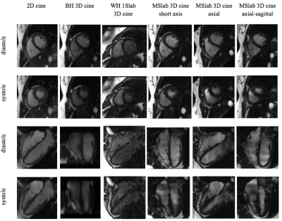
Conventional 2D cine is hindered by its needs of repeated breathhold and imaging at each individual view, while breathheld 3D cine suffers from limited slice coverage and resolution. This work developed a new 3D cine sequence with free-breathing capability for whole-heart coverage and isotropic resolution and multi-thin-slab acquisition for high blood-to-myocardium contrast.
Introduction
Conventional 2D cine MRI, although routinely used for cardiac function assessment, is subject to known limits. Its lengthy series of breathholds needed for slice position and imaging at each individual cardiac view slows down workflow and is challenging for many patients. Also, the inevitable scan-to-scan variation in breathholding position in-between short-axis slices makes the so-obtained ventricular volumes error prone. Although advances in fast imaging have enabled 3D cine in a single breathhold [1], its slice coverage remains limited to cardiac ventricles only with large slice thickness. Recent efforts have attempted to resolve cardiac motion of the entire heart in high resolution during free breathing for arbitrary offline reformatting [2, 3]. However, its whole cardiac blood pool coverage significantly compromises in-flow blood enhancement and results in low blood-myocardium contrast. In this work, we aimed to evaluate the feasibility of free-breathing whole-heart 3D cine with high contrast using multiple thin slab acquisitions.Methods
The entire heart is imaged in multiple slabs sequentially, while each slab covers a relatively thin volume to gain blood enhancement from through slab blood flow. Variable density k-t sampling was used for highly accelerated acquisition. For robustness to respiratory motion, radial vieworder with golden angle increment was employed both in [ky, kz] and along time. Respiratory gating was performed prospectively on a per cardiac phase basis during acquisition.
In reconstruction, kat ARC [4] was used to complete k-space utilizing spatiotemporal correlation. Respiratory motion was further suppressed by incorporating motion error of each k-space acquisition into the k-t reconstruction as a regularization term [5] based on simultaneously recorded bellow signal. Specifically, such regularization balances between data synthesis accuracy and motion suppression, effectively recovering k-space more reliant on data with less motion, while utilizing all acquired data for high gating efficiency.
4 healthy volunteers were scanned on a GE 1.5T 450w scanner. 3D cine imaging was performed with isotropic spatial resolution of 2.0 mm and a temporal resolution of ~50ms. 6 slabs were used to cover the entire heart with ~20 slices/slab and 9 overlapping slices to reduce venetian shading. Data were acquired in free-breathing with a gating efficiency of 60% and an acceleration factor of 8. To study the impacts of orientation to contrast and scan time, imaging was performed at 3 different orientations, 1. short-axis with slabs perpendicular to the main ventricular flow, 2. axial with the smallest phase-encoding and slice FOV and 3. axial-sagittal (an axial plane tilted in a coronal localization view to be perpendicular to the ventricular long axis) with the smallest phase encoding FOV and relative strong through slab flow. Signal in left (LV) and right ventricular (RV) blood pool and septum was measured in a 4 chamber reformat and contrast was calculated as the average blood pool signal at systole and diastole divided by the signal in septum.
