3308
Comparison between 3D white-matter-suppressed MPRAGE and 3D SPACE FLAIR in the 7T MR imaging of tuberous sclerosis complex1State Key Laboratory of Brain and Cognitive Science, Beijing MRI Center for Brain Research, Institute of Biophysics, Chinese Academy of Sciences, Beijing, China, 2University of Chinese Academy of Sciences, Beijing, China, 3Chinese PLA general hospital, Beijing, China
Synopsis
Tuberous sclerosis complex (TSC) is a multisystem, autosomal dominant disorder, which was characterized by tubers at the interface of gray and white matters. The 3D SPACE FLAIR sequence has previously been applied to detect the TSC lesions at 7T. The major limitations are its insensitivity to subtle lesions and long acquisition time. 3D white matter suppressed (WMS) MPRAGE was proposed in this study. T1-weighted 3D WMS MPRAGE suppressed the normal white matter signal selectively and made the lesions stand out. In comparison with 3D SPACE FLAIR, 3D WMS MPRAGE could detect more subtle tubers and details within reasonable time.
Introduction
Tuberous sclerosis complex (TSC) is a multisystem, autosomal dominant disorder resulted from mutations of TSC1 or TSC2 genes1. Extensive tubers usually develop at the interface of gray and white matters. Tubers exhibit abnormal cellular composition, morphology and cytoarchitecture2 and are often related to seizure. Besides T1- and T2-weighted images, 3D SPACE FLAIR was also utilized for detecting TSC tubers at 7T3. The major drawbacks of 3D SPACE FLAIR at 7T were its long acquisition time and insensitivity to early pathological changes. In this study, an alternative and fast protocol, 3D white-matter-suppressed (WMS) MPRAGE4, was proposed for delineating the TSC tubers at 7T and compared with 3D SPACE FLAIR.Methods
Thirteen patients (8-36 years old) were scanned at a 7T research system (Siemens, Erlangen, Germany) with a volume transmit/32 channel receive head coil (Nova Medical). The study was approved by the Institutional Review Board of the Beijing MRI Center for Brain Research. Besides T1- and T2-weighted images, other high-resolution imaging sequences, such as 3D WMS MPRAGE and 3D FLAIR, were also acquired to improve the TSC lesion detection capabilities. Following parameters were chosen. 3D WMS MPRAGE: field of view=230*230mm2, spatial resolution=0.72*0.72*0.72mm3, echo/repetition/inversion time=3.38/3000/580ms, scan time=5:48minutes; 3D SPACE FLAIR: field of view=206*180mm2, spatial resolution=0.8*0.8*0.8mm3, echo/repetition/inversion time=386/8200/2150ms, scan time=8:16minutes. Three experienced clinical neuroscientists and epileptologists were involved to evaluate both WMS and FLAIR images in contrast through visual inspection of TSC lesions. One healthy control was also scanned with the same WMS protocol as reference.Results
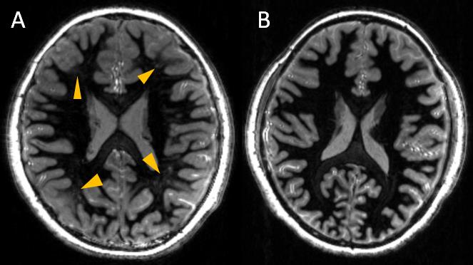
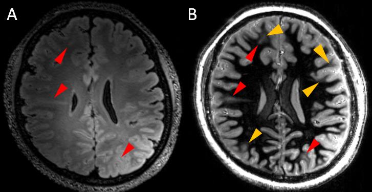
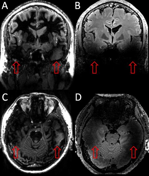
7T MRI has been performed on 13 patients (8-36 years, 7 males) with TSC and one healthy control. No adverse events occurred during the scans. As shown in Fig. 1, WM-suppressed protocol at 7T subdued the signal of white matter and revealed tubers and abnormal signals embodied in the white matter. The abnormal signals might consist of subtle tubers and transmentle signs. They demonstrated heterogeneous tissue features of white matter regions, which has striking contrast with those of the healthy control subject. 3D SPACE FLAIR with high spatial resolution at 7T demonstrated hyperintense at the lesions and showed many larger tubers. In comparison, 3D WMS MPRAGE was more sensitive to details around large tubers and subtle lesions (Fig. 2). The similar results were also found in other patients (13/13). In addition, 3D WMS MPRAGE could be attained in shorter scan time. However, the two imaging protocols both suffered from signal loss at the temporal lobes (Fig. 3) and hindered the detection of lesions in that area.Discussions and Conclusions
Accurate localization of tubers in the brains of TSC patients was very important for diagnosis and preoperative assessment. Two 7T imaging protocols, including 3D WMS MPRAGE and 3D SPACE FLAIR, were applied and compared in this study. WMS MPRAGE was achieved by setting the imaging parameters of repetition (TR) and inversion time (TI) and suppressed the normal white matter signal selectively to make tubers stand out. Unlike 3D SPACE FLAIR, T1-weighted WMS MPRAGE images reflected the different T1 values between TSC lesions and normal white matter, and could detect more subtle lesions and details. This might be due to the image blurring of 3D SPACE FLAIR or that the change of T1 value was ahead in the pathological process of TSC lesions. Thus, in term of detecting subtle tubers in the white matter, the imaging protocol of 3D WMS MPRAGE at 7T MRI system showed an advantage over the 3D SPACE FLAIR sequence.Acknowledgements
We thank Ms. Jing An (Siemens Shenzhen MR Ltd. China) for her technical assistance. This work was supported in part by the Ministry of Science and Technology of China (MOST) grants (2015CB351701, 2012CB825500), National Nature Science Foundation of China grant (91132302, 81771388), and Chinese Academy of Sciences Strategic Priority Research Program B grants (XDB02010001, XDB02050001).References
1. Crino PB, Nathanson KL, Henske EP. The tuberous sclerosis complex. N Engl J Med 2006;355:1345–1356.
2. Major P, Rakowski S, Simon M V., et al. Are cortical tubers epileptogenic? Evidence from electrocorticography. Epilepsia. 2009; 50:147–154.
3. Chalifoux JR, Perry N, Katz JS, et al. The ability of high field strength 7-T magnetic resonance imaging to reveal previously uncharacterized brain lesions in patients with tuberous sclerosis complex. J Neurosurg Pediatr. 2013; 11:268–273.
4. Tourdias T, Saranathan M, Levesque IR, Su J, Rutt BK. Visualization of intra-thalamic nuclei with optimized white-matter-nulled MPRAGE at 7T. Neuroimage. 2014;84:534–545.
Figures