3306
Evaluation of Diffusion-Weighted Imaging of the Macaque Brains Using Readout-Segmented EPI at 7TPinyi Wang1,2, Jialu Zhang1,2,3, Meizhen Qian1,4, Yi Sun5, Dingxin Wang3, and Xiaotong Zhang1,2,6
1Interdisciplinary Institute of Neuroscience and Technology, Qiushi Academy for Advanced Studies, Zhejiang University, Hangzhou, China, 2College of Biomedical Engineering & Instrument Science, Zhejiang University, Hangzhou, China, 3Center for Magnetic Resonance Research, University of Minnesota, Minneapolis, MN, United States, 4School of Medicine, Zhejiang University, Hangzhou, China, 5MR Collaboration NE Asia, Siemens Healthcare, Shanghai, China, 6Key Laboratory for Biomedical Engineering of Ministry of Education, Zhejiang University, Hangzhou, China
Synopsis
Diffusion-weighted imaging (DWI) is an important tool for clinical diagnosis and neuroscience research. To evaluate DWI with higher spatial resolution and with reduced image distortion at higher field strengths, we conducted macaque brain imaging at 7T. Our results suggest that readout-segmented EPI (rsEPI) has reduced image distortion, high MR signal and image contrast. It is believed that the rsEPI can effectively benefit DWI for macaque brain researches at 7T.
Introduction
Diffusion-weighted imaging (DWI) is an important tool for clinical diagnosis and neuroscience research.1 The single-shot echo planar imaging (ssEPI) sequences have been widely used; however, they suffer from well-known motion-related artifacts as well as the subtle T2* blurring due to a significant T2* decay of the signal and phase shift accumulation. Therefore, these problems hinder them from achieving higher spatial resolution especially at high field strengths. A feasible approach to overcome such problems is to use diffusion-weighted, readout-segmented EPI (rsEPI) with 2-dimensional navigator correction,2,3 which has lower susceptibility distortion, reduced T2* blurring and a robust correction for motion-induced phase artifact. In the current study, through evaluating image distortion, signal strength and contrast, we provide a DWI evaluation by employing the rsEPI for in vivo macaque brain imaging over a 7T human MR scanner.Methods
All measurements were performed on a 7T research MR scanner (Siemens Healthcare, Erlangen, Germany) equipped with a 1Tx/28Rx QED knee coil (Mayfield Village, OH, USA). Images were acquired over a female macaque (2-year old, 3.5kg) that was maintained and anesthetized by 1.5~2% isoflurane – all procedures were in accordance with NIH standards and approved by our Institutional Animal Care Committee. The monkey was placed in the sphinx position with its head centered within the knee coil. T2w TSE sequence were obtained in the same slice position as a reference for distortion evaluation (TR/TE 9000/36 ms; FOV 128×128 mm2; matrix 128×128). Prototype sequence RESOLVE4,5 was utilized for rsEPI, and DW images were acquired at 1×1×1 mm3 resolution with an acceleration rate of 2 and b-values of 0, 500 and 1000 s/mm2. The following parameter were used for all rsEPI-DWIs: matrix 96×96, FOV 96 mm; TR 7300 ms, acceleration rate 2 along A-P phase encoding direction, 2 averages, bandwidth of 789 Hz/pixel. We evaluated DW image with a monopolar scheme but increased readout segmented numbers of 7, 13, and 19 with the echo spacing of 0.4 ms, 0.34 ms, and 0.32 ms, and the scan time of 13’44”, 25’04”, and 35’31”, respectively. We also acquired DW images with 7 shots but for contrast-comparison purpose by using the bipolar and monopolar schemes.Results and Discussion
Figure 1 shows rsEPI images at b = 0 s/mm2 acquired at 7T in 17'29" with a voxel size of 0.7×0.7×1 mm3. T2-weighted images by TSE were provided as a reference for distortion evaluation. The squares in Figure 1 were drawn on the same slice position to enclose the same region of interest for geometric distortion comparison. The rsEPI DWIs showed a certain level of image distortion in regions with air-tissue interfaces, such as located at the paranasal sinus, the nasal cavity and the ear cavity. Figure 2 shows a comparisons of image distortion with increased readout segmented number for rsEPI with a monopolar scheme and 1 mm isotropic resolution (25 slices). Increasing shots number with rsEPI leads to longer scan time but shorter readout echo spacing, which of the latter is supposed to alleviate image distortions due to the susceptibility effect2,3. However, the DW images distortion observed in Figure 2 were not apparently reduced with increased shot number. Figure 3 shows a comparison of 1mm isotropic DWIs (54 slices) between a bipolar scheme (left) and a monopolar (right) with 7-shot rsEPI-DWI acquired at 7T. The monopolar scheme is more sensitive to eddy currents induced by the diffusion gradient. Therefore, the bipolar scheme is usually used to avoid eddy currents that can arise from switching the diffusion gradient rather than a monopolar6,7; however, our results indicates that the monopolar scheme had slight distortion and higher signal intensity/contrast as compared to the bipolar scheme.Conclusion
The results shown in this study demonstrate that, for in vivo macaque brain imaging at 7T, rsEPI makes it possible to perform DW imaging with multiple readout segmentation and 1mm isotropic resolution with restrained images distortion and satisfactory MR signal quality. However, distortion in rsEPI-DWI were not apparently reduced with increased shots number (7, 13, and 19). In addition, the DW images with a monopolar scheme outperforms the bipolar scheme in signal intensity and contrast. Overall, rsEPI-based diffusion imaging can play an important role in high-resolution macaque brain research at 7T.Acknowledgements
We would like to thank Mr. Guohua Xu, Bin Xu, and Dengfeng Zhou for animal handling. This work was supported in part by National Natural Science Foundation of China (81701774, 61771423) and Fundamental Research Funds for the Central Universities (2016QN81018).References
1. Mansfield P, Pykett I L. Biological and medical imaging by NMR. 1978[J]. Journal of Magnetic Resonance, 2011, 213(2):513. 2. Pipe J G. Motion correction with PROPELLER MRI: Application to head motion and free‐breathing cardiac imaging[J]. Magnetic Resonance in Medicine, 1999, 42(5):963. 3. Pipe JG, Farthing VG, Forbes KP. Multishot diffusion-weighted FSE using PROPELLER MRI.[J]. Magnetic Resonance in Medicine, 2002, 47(1):42. 4. Robson M D, Anderson A W, Gore J C. Diffusion-weighted multiple shot echo planar imaging of humans without navigation[J]. Magnetic Resonance in Medicine, 1997, 38(1):82. 5. Miller K L, Pauly J M. Nonlinear phase correction for navigated diffusion imaging[J]. Magnetic Resonance in Medicine, 2003, 50(2):343. 6. Andrew L A P D, Tsuruda J S, Parker D L. Elimination of eddy current artifacts in diffusion‐weighted echo‐planar images: The use of bipolar gradients[J]. Magnetic Resonance in Medicine, 1997, 38(6):1016-21. 7. Reese T G, Heid O, Weisskoff R M, et al. Reduction of eddy‐current‐induced distortion in diffusion MRI using a twice‐refocused spin echo[J]. Magnetic Resonance in Medicine, 2003, 49(1):177.Figures
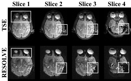
Fig.1 Examples of rsEPI
images at b = 0 s/mm2 with 7 shots and a monopolar scheme acquired
at 7T in 17'29" min (voxel size of 0.7×0.7×1mm3). T2-weighted TSE
were provided as a reference for distortion evaluation. The squares were drawn
on the same slice position to enclose the same regions of interest for
geometric distortion comparison.
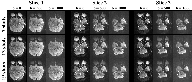
Fig.2 Comparison of 1×1×1mm resolution DWIs between
7 (an echo spacing of 0.4ms), 13 (an echo spacing of 0.34ms), 19 (an echo
spacing of 0.32ms) readout segmented rsEPI in the macaque brain at b-value of 0, 500 and 1000 s/mm2
with 25 slices acquired at 7T.
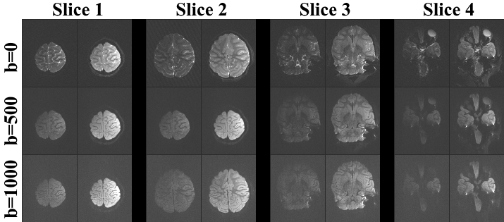
Fig.3 Comparison of 1 mm isotropic
resolution DWIs between a bipolar scheme (left) and a monopolar (right) 7
readout segmented rsEPI-DWI in the macaque brain at b-value of 0, 500 and 1000 s/mm2 with 54 slices acquired
at 7T.