3303
Probing Activated Regions in Rat’s Olfactory System by Odor Stimulations through Manganese Enhanced MRI (MEMRI) at 7TBin Zhang1, Qunchen Yuan1, Zhen Qin1, Liujing Zhuang1, Ping Wang1,2, and Xiaotong Zhang1,2
1Key Laboratory for Biomedical Engineering of Ministry of Education, College of Biomedical Engineering & Instrument Science, Zhejiang University, Hangzhou, China, 2Interdisciplinary Institute of Neuroscience and Technology, Qiushi Academy for Advanced Studies, Zhejiang University, Hangzhou, China
Synopsis
In vivo bioelectronic nose utilizes mammalian olfactory system as a means of odor detection and discrimination. However, electrode localization during implantation relies on the researcher’s experiences, resulting in an unreliable success rate. The goal of this research is to determine the optimal electrode implantation positions for electrophysiological recordings by using the technique of manganese enhanced MRI (MEMRI) at 7T. A small dose of manganese ion was delivered into the rat’s right naris and an odor was delivered to its nose during MRI scanning. With the MRI data, the region activated by the specific odor can be identified in the OB.
Introduction
In vivo bioelectronic nose has been developed based on brain-computer interface (BCI) and neural decoding techniques, utilizing mammalian olfactory system as a means of odor detection and discrimination. By implanting electrodes into animal’s olfactory bulb (OB) and recording electrophysiological signals, odor information can be extracted.1,2 Yet the success rate of in vivo bioelectronic nose is not satisfactory since the implantation site is mostly based on experience and the recorded neurons may not be responsive to the designated odorant. The goal of this research is to determine the optimal electrode implantation positions for electrophysiological recordings by using the technique of manganese enhanced MRI (MEMRI). MEMRI uses exogenous manganese ions (Mn2+) as the contrast agent based on the paramagnetism and calcium ion (Ca2+) similarity of the manganese ions.3 When the olfactory sensory neurons (OSNs) in the epithelium are activated by odorants and the Ca2+ channels are opened, the Mn2+ enters the olfactory system through the Ca2+ channels and accumulates in the olfactory pathway, then Mn2+ accumulation can be probed and localized through conventional MRI technique such as T1w imaging.Methods
All procedures were in accordance with NIH standards and with approval of our University Institutional Animal Care Committee. Adult Sprague-Dawley Rats (200–250 g) were anesthetized with an intraperitoneal injection of chloral hydras (10%, 0.4 ml/kg), 20 μL MnCl2 (400 mM) was dropped into each rat’s right naris using a pipette (by four times, at different depths of the naris each time). The rat was kept supine for 10 minutes and odorant stimulation was conducted simultaneously by placing a small glass dish containing 0.5 mL odorant (10-3 mM, diluted by mineral oil) in front of the rat’s nose. The MRI measurements were performed on a 7T research scanner (Siemens Healthcare, Erlangen, Germany), and the rat was fixed on an MRI-compatible stereotaxic apparatus and placed in the prone position inside the MRI bore. A custom-built 1Tx/5Rx surface array4 was mounted above the rat’s head to image its entire brain, and the odorant was exposed to the rat throughout the entire scan. With every 30 min time interval, axial T1-weighted 3D FLASH images were obtained with an acceleration rate of 2 along F-H phase encoding direction (TE 5.84ms, TR 435ms, matrix size 320×320, FOV 32mm, slice thickness 0.5mm, FA 5, 12 averages, scan time 14’54”).Results
30 min after Mn2+ administration, the MRI signal intensity of some regions in the right olfactory epithelium (OE) decreased (darker in the images), but no obvious change was observed in the OB, suggesting the Mn2+ started to accumulate in the OE but hadn’t arrived in the OB yet (figure not shown). In about 1 hour, Mn2+ could be seen in the right OB (Fig.1a), resulting a significant decrease of signal intensity (P < 0.001) (fig 2). With the accumulation of Mn2+, the signal intensity of the red circles in Fig.1 keeps decreasing (Fig.2). Fig.3 shows the slice images of three rats (Rat A, B and C) from three trials 1 h after Mn2+ administration. Rat A, B and C were stimulated by diacetyl, isoamyl acetate and N-pentanal respectively. Different odorants induced different Mn2+ accumulation regions in the OB. The induced region of Rat A is in most of the lateral OB, and soamyl acetate activated some parts of the ventral and lateral OB, whereas N-pentanal activated ventral lateral anterior OB. Note that these activated regions were partially overlapped.Discussion
During different odorant stimulation trials, OSNs responded to different odorants, so Mn2+ entered different neurons and accumulated in different regions in the OB. The overlapping of the odor-induced regions may be a result of cross reaction of OSNs with odorants.1 An odorant incites various responses of different OSNs, which project to multiple glomeruli in the OB; on the other hand, the same OSNs projecting to the same glomerulus can be activated by many odorants.Conclusion
Taking the advantage of MEMRI, this research has shown the olfactory pathway, verified the cross reaction of OSNs/glomeruli with odorants and found topographic patterns of responsiveness to odorants in the rat’s OB. Moreover, this study helps determine the electrode implantation sites for electrophysiological recordings, which would effectively improve the performance of in vivo bioelectronic nose.Acknowledgements
This work was supported in part by National Natural Science Foundation of China (81701774, 61771423), Major International Cooperation Project of Natural Science Foundation of China (61320106002, 31661143030), and Fundamental Research Funds for the Central Universities (2016QN81018).References
1. Zhuang L, Guo T, Cao D, et al. Detection and classification of natural odors with an in vivo bioelectronic nose.[J]. Biosensors & Bioelectronics, 2015, 67:694-699. 2. Zhuang L, Hu N, Tian F, et al. A high-sensitive detection method for carvone odor by implanted electrodes in rat olfactory bulb[J]. Chinese Science Bulletin, 2014, 3. Pautler R G, Koretsky A P. Tracing odor-induced activation in the olfactory bulbs of mice using manganese-enhanced magnetic resonance imaging.[J]. Neuroimage, 2002, 16(2):441. 4. Zhang X, Gao Y, Adriany G. A Surface Loop Array for Small Animal Imaging at 7T. Proceedings of ISMRM, 2018, submitted.Figures
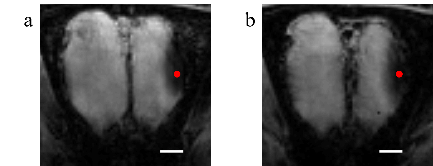
Fig.1
T1w maps of rat’s olfactory system changed over time after Mn2+
administration and diacetyl stimulation. (a) 1 hour after Mn2+ administration,
and (b) 2 hour after Mn2+ administration. (Scale bar = 1 mm.)
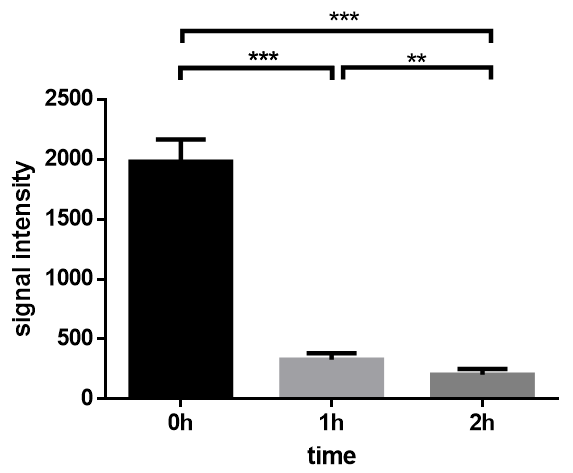
Fig.2
MRI signal intensities at the same position (the red circles in Fig.1) of rats’
OBs along time after Mn2+ administration and diacetyl stimulation.
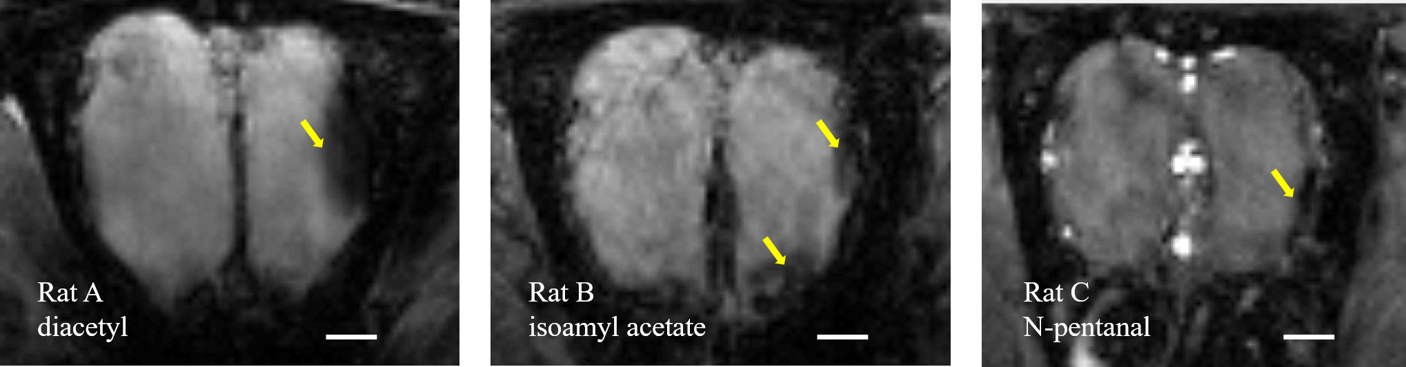
Fig.3
Mn2+ accumulation regions varied when rats were stimulated by
different odors. (Scale bar = 1 mm.)