3302
Spinal Cord Gray Matter Myeloarchitecture Revealed by 7T Magnetization Transfer Imaging1Translational and Molecular Imaging Institute, Icahn School of Medicine at Mount Sinai, New York, NY, United States, 2Department of Radiology, Icahn School of Medicine at Mount Sinai, New York, NY, United States, 3Graduate School of Biomedical Sciences, Icahn School of Medicine at Mount Sinai, New York, NY, United States, 4Department of Neuroscience, Icahn School of Medicine at Mount Sinai, New York, NY, United States
Synopsis
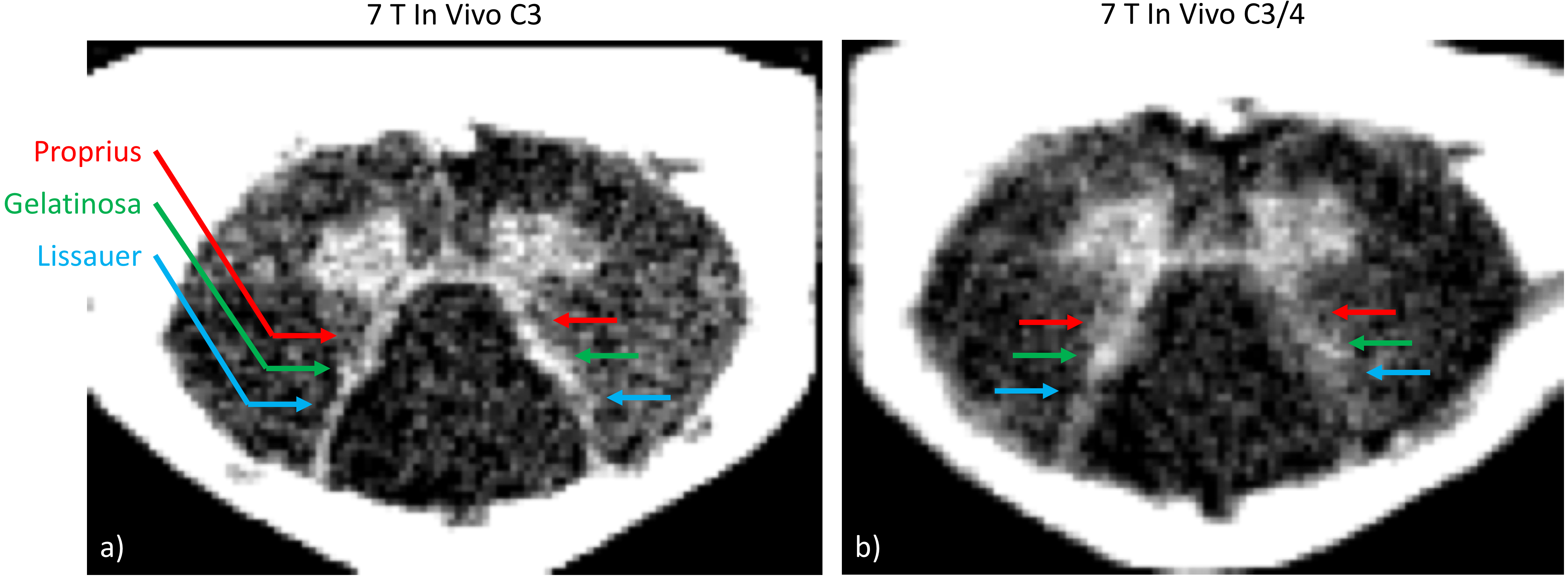
The human spinal cord gray matter contains multiple nuclei and laminae, which are not usually distinguishable in MRI at conventional field strengths. The higher signal-to-noise ratio achievable at 7T provides the increased spatial resolution necessary to resolve these very small gray matter features. Using a wrap-around brainstem/cervical spinal cord RF coil at 7T and a magnetization transfer-prepared multi-echo gradient echo pulse sequence, we resolved three differentially myelinated dorsal horn gray matter structures: the dorsolateral fasciculus, substantia gelatinosa, and nucleus proprius in a single subject at 150-µm in-plane resolution.
Introduction
Cervical spinal cord MRI has important applications in the study of neurological disorders such as multiple sclerosis, cervical spondylotic myelopathy, and spinal cord injury. At 3T, where spatial resolution is limited, spinal cord cross-sectional area is a commonly-calculated biomarker of spinal cord atrophy1. The higher signal-to-noise ratio achievable at ultra-high field (7T) can provide the increased spatial resolution necessary to resolve and investigate sub-regions of the spinal cord, such as individual white matter tracts or gray matter nuclei. The achievable signal-to-noise ratio, and ultimately the achievable image resolution, is also strongly dependent on radiofrequency hardware. Taso et al. have recently presented a parcellation of the cervical spinal cord white and gray matter by k-means clustering based on data acquired from eight subjects using an 8-channel transceiver array at 7T2. Using a 22-channel receive, 4-channel transmit, two-panel RF coil at 7T3 and a magnetization transfer-prepared multi-echo gradient echo pulse sequence, we have acquired images in a single subject at 150-µm in-plane resolution in which myelin contrast differentiates the dorsolateral fasciculus of Lissauer, the substantia gelatinosa of Rolando (Rexed lamina II), and the nucleus proprius (Rexed laminae III and IV) in the dorsal horn gray matter.Methods
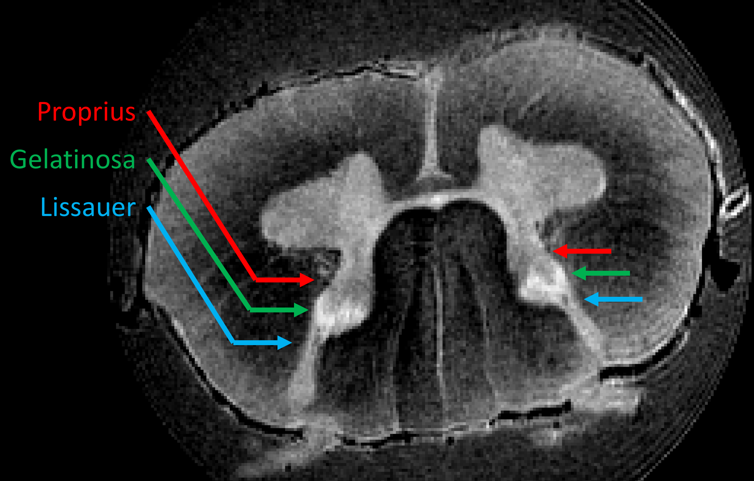
The 22-channel receive, 4-channel transmit, two-panel RF coil described in Zhang et al.3 was interfaced to a 7T human whole-body MRI scanner (Magnetom, Siemens). Magnetization transfer-prepared multi-echo spoiled GRE images were acquired at the level of the C3 vertebral body and the C3/C4 intervertebral disc, at 0.15 x 0.15 x 1.5 mm3 resolution in a single slice. TR/TE1/TE2/TE3 = 325/5.6/15.4/25.2ms, α=45°, and Gaussian magnetization transfer preparation pulses of 720° were applied at 1750Hz off-resonance. This acquisition was repeated three and five times at C3 and C3-C4, respectively. The resulting multi-echo images from each acquisition were co-registered in two dimensions based on the first-echo image using ANTs4, and the echoes of repeated acquisitions were summed individually. The three echoes were then combined by root-sum-of-squares and smoothed with a Gaussian kernel with sigma=0.5 pixels. An ex vivo image of a human cervical spinal cord was also acquired at 9.4T using a 3D RARE sequence at 63 x 63 x 250 µm3 resolution for comparison.Results
Three differentially myelinated features are visible in vivo (Figure 1): the myelinated dorsolateral fasciculus of Lissauer, the unmyelinated substantia gelatinosa of Rolando (Rexed lamina II), and the moderately myelinated nucleus proprius (Rexed laminae III and IV); all are spatially consistent with an ultra-high-resolution ex vivo 9.4 T RARE image (Figure 2).Discussion
Multi-echo gradient echo images with combined proton-density and T2* contrasts have been shown to provide excellent gray/white matter contrast in the spinal cord3. The addition of magnetization-transfer contrast presumably accentuates the contrast between differentially myelinated structures5, although the relative contributions of T2* and magnetization-transfer contrasts need further investigation.
The dorsolateral fasciculus of Lissauer and the substantia gelatinosa of Rolando, are both components of the nociceptive system. Thinly-myelinated sensory axons conveying pain signals enter the spinal cord, ascend or descend up to two spinal levels in the dorsolateral fasciculus of Lissauer, and then synapse in the substantia gelatinosa of Rolando. The substantia gelatinosa, containing many neuronal cell bodies and very few myelinated axons, is an important site for modulation of nociception via opioid receptors. Injury or degradation of these structures may have significant effects on sensation and nociception.
Conclusion
Resolving myeloarchitectural features in the dorsal horn of the spinal cord gray matter not only demonstrates the advantage of ultra-high field MRI with optimized radiofrequency hardware, but also paves the way to study these small structures and their role in sensory processing.Acknowledgements
No acknowledgement found.References
[1] De Leener B, Kadoury S, Cohen-Adad J. Robust, accurate and fast automatic segmentation of the spinal cord. NeuroImage 2014;98:528-536.
[2] Taso M, Massire A, Besson P, Le Troter A, Guye M, Ranjeva J-P, Callot V. Towards in vivo spinal cord cyto- and myelo-architecture deciphering using multi-modal MRI parcellation at 7T. Proc. ISMRM 2017 #0018.
[3] Zhang B, Seifert AC, Kim JW, Borrello J, Xu J. 7 Tesla 22-channel wrap-around coil array for cervical spinal cord and brainstem imaging. Magn. Res. Med. 2017;78:1623-1634.
[4] Avants BB, Tustison NJ, Song G, Cook PA, Klein A, Gee JC. A reproducible evaluation of ANTs similarity metric performance in brain image registration. NeuroImage 2011;54:2033-2044.
[5] Henkelman RM, Stanisz GJ, Graham SJ. Magnetization transfer in MRI: a review. NMR Biomed 2001;14:57-64.
Figures