3287
Individual patient-specific quantification of the network effects of lesions and diffuse disconnections with diffusion MRI1Neurology, Max Planck Institute for Human Cognitive and Brain Sciences, Leipzig, Germany, 2Cerebral Imaging Center, Douglas Mental Health University Institute, McGill University, Montreal, QC, Canada, 3Montreal Neurological Institute, McGill University, Montreal, QC, Canada, 4Spinoza Centre for Neuroimaging. Netherlands Institute for Neuroscience, Amsterdam, Netherlands
Synopsis
Even with its crucial role as the backbone of communication within the brain and susceptibility to damage from diseases such as stroke and multiple sclerosis, the white-matter connectional architecture of the human brain has been largely ignored. To address this, we have developed a whole-brain model for the quantification of white-matter connectional anatomy – the Tractography-based Lesion Assessment Standard (TractLAS) – which provides a complete connectional network description of the human brain. It can be used to quantify the impact of lesions on the connectivity of the brain within individuals, providing clinically useful information for individualised care.
Introduction
With an estimated five million new stroke survivors every year 1 and a rapidly aging population suffering from multiple sclerosis, white-matter hyperintensities, and other diseases of presumed vascular origin 2 that affect white matter and contribute to cognitive decline 3, it is crucial that we understand how white-matter damage affects the brain and behaviour so that we can develop targeted interventions to improve recovery and ameliorate decline. Current MRI techniques for assessing the impact of brain damage/lesions consider only location, type, and extent, while ignoring how the affected region is or was connected to the rest of the brain. A comprehensive network-based perspective can provide a more complete characterisation of the structural impact of lesions that can be used to better understand functional deficits and recovery trajectories4,5. To address these issues and provide individual patient-specific information for individualised monitoring and care, we created the TractLAS to enable the quantification of lesions and connectional disruptions on brain connectivity.Method
80 (40 female) unrelated healthy young adults from the Human Connectome Project S900 data release (1.25 isotropic dMRI, 90 directions per shell, b=1000/2000/3000 s/mm2) were used to create the tractography atlas. Images were processed with MRtrix6 to generate fODFs and perform whole-brain anatomically-constrained tractography. Tractography was performed between all grey-matter regions. To preserve the information about the connectional architecture necessary for assessing the impact of lesions, region-to-region connectivity matrices were extracted to estimate connectivity between all grey-matter and white-matter regions within the brain in each individual.Results
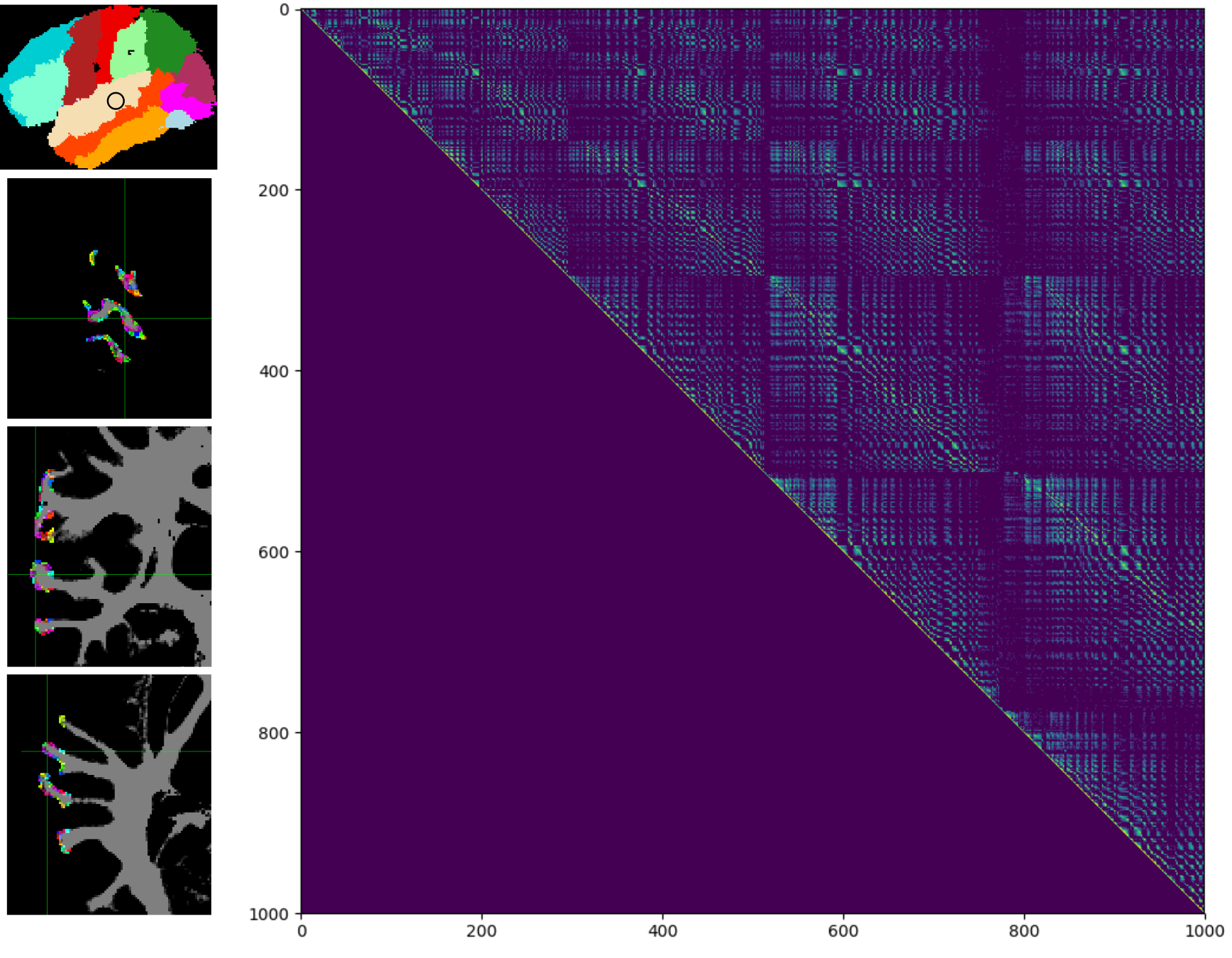
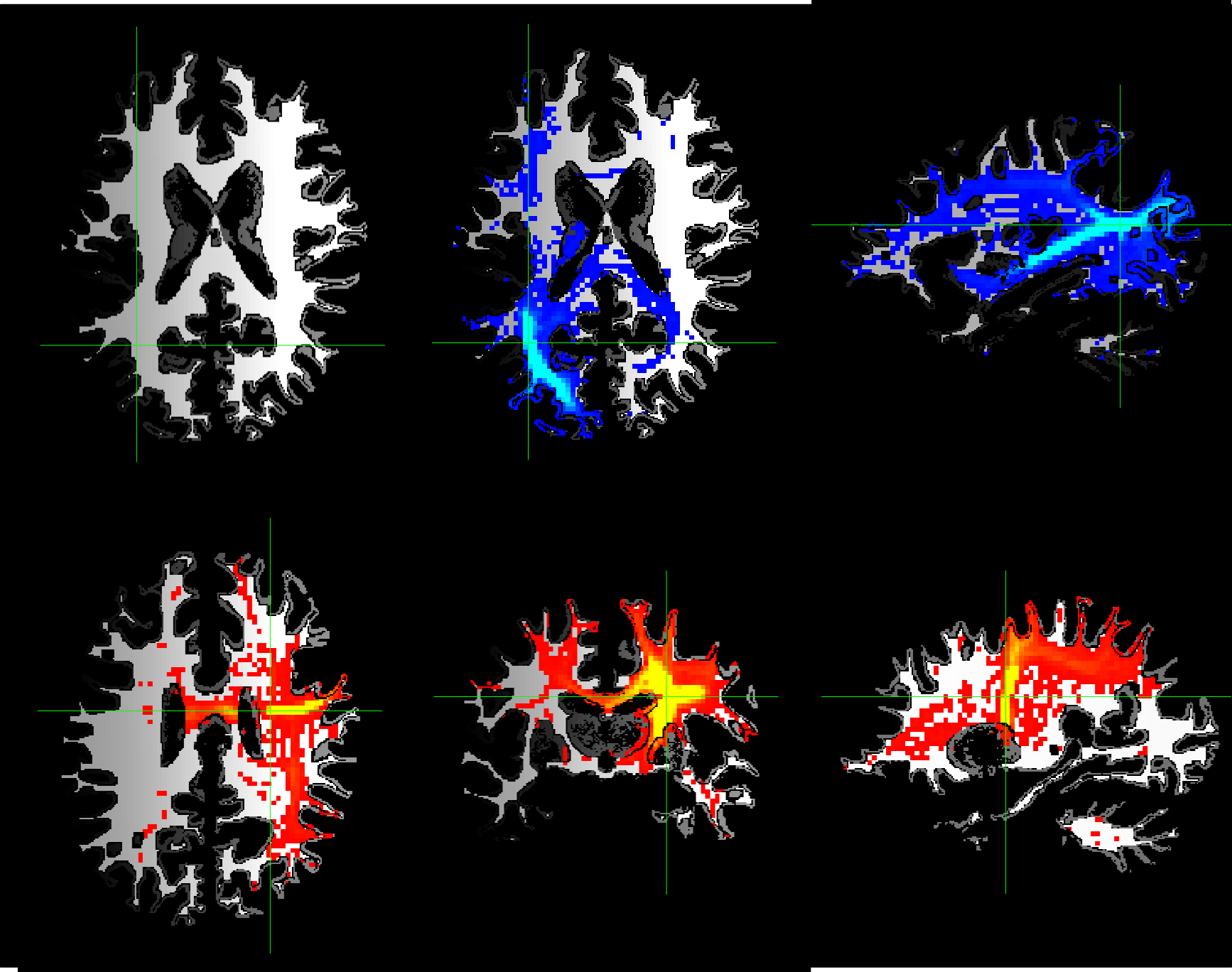
We created a tractography standard consisting of region-to-region connectivity matrices for all white- and grey-matter regions in the brain. Our model retains the spatial information of white-matter connectivity in order to assess the impact of spatially disparate lesions, which results in extremely large connectivity matrices (grey-grey, white-white, grey-white) that necessitated the creation of custom processing and analysis tools. Our standard provides an extremely high level of connectional granularity, computing connectivity between individual voxels or sets of voxels rather than grossly defined atlas regions (Figure 1). Co-registered lesion maps are transformed into the matrix space, predicted change in probabilistic connectivity is computed and can be transformed back into a standard space for visualisation (Figure 2). This is performed on an individual level, thus individual patient lesions can be (a) assessed for the grey- and white-matter regions that they probabilistically connect to and (b) have disconnection quantified relative to a healthy young adult standard.Discussion and Conclusions
We have created a standard model of white-matter connectional anatomy that can be used to both assess and monitor the individual effects of lesions (stroke, multiple sclerosis, white-matter hyperintensities) during disease progression and rehabilitation. Our model allows the generation of individual structural disconnection maps that can be used to quickly identify affected white-matter tracts and connected regions, and relate them to clinical symptoms and behavioral deficits. This work expands upon the established standard lesion-deficit paradigm to assess disconnectivity throughout the whole brain with a network perspective.Acknowledgements
No acknowledgement found.References
1. Wahlund, L. O. et al. A New Rating Scale for Age-Related White Matter Changes Applicable to MRI and CT. Stroke 32, 1318–1322 (2001).
2. Bates, E. et al. Voxel-based lesion–symptom mapping. Nat Neurosci 6, 448–450 (2003).
3. Lampe, L et al., Lesion location matters: effects of white matter hyperintensities on gray matter and cognition in the healthy elderly, JCBFM (in press, 2017).
4. Ovadia-Caro, S. et al. Longitudinal effects of lesions on functional networks after stroke. J Cereb Blood Flow Metab 33, 1279–1285 (2013).
5. Kuceyeski, A et al. (2013) The Network Modification (NeMo) Tool: Elucidating the Effect of White Matter Integrity Changes on Cortical and Subcortical Structural Connectivity. Brain Connect 3:451–463.
6. Tournier, J-D et al., (2012) MRtrix: Diffusion tractography in crossing fiber regions. Int J Imaging Syst Technol 22:53–66.
Figures