3286
Short-term Visual Experience Increases Intrinsic Brain Connectivity Within Ventral Visual Pathway1Department of Medical Imaging, First Affiliated Hospital of Medical College, Xi’an Jiaotong University, Xi'an, China
Synopsis
Medical imaging interpretation fundamentally lies in radiologists’ exceptional visual recognition skill, which enables the identification of pathological regions to render diagnosis1. Such expertise is obtained through training across review hundreds of cases2, facilitated by the plastic changes in the central visual system3. Specifically, ventral visual pathway (VTP) is responsible for visual object recognition, i.e. the fine-grained visual information processing. Visual information processing in the adult human brain is highly malleable with neural processing adapting to incoming information4. The plastic changes in the VTP in response to visual recognition tasks are well studied5. While we propose that the information embedded in the intrinsic brain activity, as revealed in the resting data, is also important6. Therefore, in the current study, we investigate how visual experience, i.e. short-term radiological training, modulates brain activity in the VTP under task-free state in the resting brain using ICA.
Introduction, Methods, Results, and Conclusion
Introduction: Medical imaging interpretation fundamentally lies in radiologists’ exceptional visual recognition skill, which enables the identification of pathological regions to render diagnosis1. Such expertise is obtained through training across review hundreds of cases2, facilitated by the plastic changes in the central visual system3. Specifically, ventral visual pathway (VTP) is responsible for visual object recognition, i.e. the fine-grained visual information processing. Visual information processing in the adult human brain is highly malleable with neural processing adapting to incoming information4. The plastic changes in the VTP in response to visual recognition tasks are well studied5. While we propose that the information embedded in the intrinsic brain activity, as revealed in the resting data, is also important6. Therefore, in the current study, we investigate how visual experience, i.e. short-term radiological training, modulates brain activity in the VTP under task-free state in the resting brain using ICA.
Methods: A group of novice radiography students (n= 21), namely the intern radiologists group (IRG), and a group of matched healthy controls (n=21), namely the normal control group (NCG), were recruited. All the subjects gave informed consent and had norm and corrected-to-normal vision when participating tests outside the scanner and went through behavioral prescreening test and MRI scanning. The behavioral test was used to evaluate the visual recognition performance in radiological domain. The resting images were obtained with an echo-planar imaging (EPI) sequence on a 3-Telsa MRI system. The axial 3D T1-weighted images were obtained to exclude subjects with prominent neurological deficits. Data preprocessing steps include slice-timing, realignment, co-registration, normalization, smooth, detrend and filtering (0.01-0.08Hz) using SPM12. ICA analysis was carried out using spatial ICA as implemented in the GIFTv4.0a toolbox. ICA was performed using Informax algorithm. Using Spatial correlation criteria, we used the template of the occipital and the temporal (obtained from the WFUpick_atlas to identify the independent component that we are interested in. The group difference in the brain activity of the VTP was investigated using two sample t-test in SPM12 (t-test, p<0.05, Alphasim corrected). Furthermore, the voxel-wise whole brain correlation analysis between the brain measurementand with the behavioral measurement was conducted ( p<0.05, Alphasim corrected).
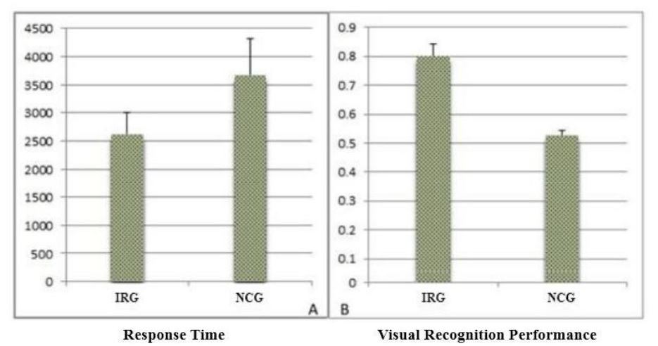
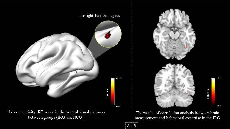
Results: The IRG significantly outperformed the NCG in behavioral expertise test (Mann-Whitney test, p<0.001), the behavioral performance can be seen in figure 1. Significantly higher brain connectivity strength in the right fusiform gyrus (FG), (p<0.05, Alphasim correted, peak voxel coordinates 41 - 59 -17) was found in the IRG, as shown in figure 2A. Additionally, connectivity strength of the right FG fusiform gyrus correlated with the intern radiologists’ behavioral expertise (corrected for multiple comparisons), as shown in figure 2B.
Conclusion: Our results have shown that short-term expertise modulates short-term radiological experience alters intern radiologists’ connectivity strength within the VTP in the brain regions, i.e. the right FG, that are likely to be engaged in their daily practice. FG is involved in fine-feature visual recognition in perceptual expertise studiesfrom different domains7. We suggest that our study may shed light on the understanding the development of visual recognition skills by illustrating that the information processing efficiency in the critical node of higher visual cortex increases8, as indexed by the increased connectivity strength, as visual expertise develops . Our current provides the one of the first evidence of neural substrates of object recognition expertise under resting state.
Acknowledgements
No acknowledgement found.References
1. Krupinski EA. Current perspectives in medical image perception. Atten Percept Psychophys. 2010; 72(5):1205-1217.
2. Nodine CF, Mello-Thoms C, Kundel HL, et al. Time course of perception and decision making during mammographic interpretation. AJR Am J Roentgenol. 2002;179(4):917-923.
3. Fahle M. Perceptual learning: a case for early selection. J Vis. 2004;4(10):879-890.
4. Op de Beeck HP, Baker C I. The neural basis of visual object learning. Trends Cogn Sci. 2010; 14(1):22-30.
5. Bilalic M, Grottenthaler T, Nagele T, et al. The Faces in Radiological Images: Fusiform Face Area Supports Radiological Expertise. Cereb Cortex. 2016;26(3):1004-1014.
6. Miall R, Robertson E. Functional Imaging: Is the Resting Brain Resting? Curr Biol. 2006;16(23):998-1000.
7. Bilalic M, Langner R, Ulrich R, et al. Many faces of expertise: fusiform face area in chess experts and novices. J Neurosci. 2011;31(28):10206-10214.
8. Fenske MJ, Aminoff E, Gronau N, et al. Top-down facilitation of visual object recognition: object-based and context-based contributions. Prog Brain Res. 2006;155:3-21.
Figures