3283
Modulation of cortico-subcortical functional connectivity occurs after symptomatic treatment of fatigue in patients with multiple sclerosisPaola Valsasina1, Bruno Colombo2, Paolo Preziosa1,2, Vittorio Martinelli2, Andrea Falini3, Giancarlo Comi2, Massimo Filippi1,2, and Maria A. Rocca1,2
1Neuroimaging Research Unit, INSPE, Division of Neuroscience, San Raffaele Scientific Institute, Vita-Salute San Raffaele University, Milan, Italy, 2Department of Neurology, San Raffaele Scientific Institute, Vita-Salute San Raffaele University, Milan, Italy, 3Department of Neuroradiology, San Raffaele Scientific Institute, Vita-Salute San Raffaele University, Milan, Italy
Synopsis
In this study, 45 fatigued patients with multiple sclerosis (MS) were randomly assigned to undergo treatment with fampridine, amantadine or placebo and underwent clinical, neuropsychological and 3T resting state (RS) functional MRI at baseline and after four weeks of treatment. We found that treatment with fampridine (and, to a lesser extent, with amantadine) ameliorates fatigue in MS. Concomitant increase of RS functional connectivity (FC) in inferior frontal and parietal cortical regions, and decrease of abnormally high intra-thalamic FC were detected, suggesting an improved regulation of cortico-subcortical functional circuits.
Introduction.
Fatigue affects a large proportion of patients with multiple sclerosis [1] and has been associated with functional abnormalities of cortico-subcortical circuits, involving fronto-parietal regions and the basal ganglia [2]. Several medications have been tested to treat fatigue in MS; however, data on their efficacy are still controversial [3]. Aim of this study was to investigate longitudinal changes of brain resting state (RS) functional connectivity (FC) in MS patients with fatigue undergoing different symptomatic treatments for this symptom.Methods.
Forty-five fatigued MS patients were randomly, blindly assigned to undergo treatment with fampridine (n=15), amantadine (n=15) or placebo (n=15) and underwent clinical, neuropsychological (including fatigue assessment) and 3T RS functional MRI at baseline (T0) and after four weeks (W4) of treatment. Fifteen matched healthy controls were acquired twice. RS FC analysis of the main brain functional networks was performed using independent component analysis [4] and statistical parametric mapping.Results.
At T0, compared with controls, MS patients showed increased intra-thalamic RS FC and abnormal fronto-parietal RS FC of several cortical networks. At W4, significantly decreased global, physical and cognitive (p=0.001/0.003/0.01) modified fatigue impact scale (MFIS) scores were found in fampridine patients and, to a lesser extent, in amantadine patients (cognitive and psycho-social MFIS, p=0.04). Placebo patients also showed improved global, physical and psycho-social MFIS (p=0.02/0.01/0.02). At W4, fampridine patients showed increased RS FC of the bilateral precuneus in the default mode and executive control networks, and increased RS FC of the right inferior frontal gyrus in the salience and fronto-parietal attention networks (Figure 1). At W4, small clusters of increased RS FC in frontal regions and decreased RS FC in temporo-parietal regions were detected in placebo and amantadine patients. A significant decrease over time of intra-thalamic and subcortical RS FC was found in fampridine and amantadine patients (Figure 2).Conclusions.
Treatment with fampridine (and, to a lesser extent, with amantadine) ameliorates fatigue in MS patients. Concomitant modifications of RS FC suggest an improved regulation of cortico-subcortical functional circuits.Acknowledgements
This work has been partially supported by a grant from Italian Ministry of Healthy (GR-2008-1138784).References
[1] Krupp LB et al., Arch Neurol 1988;45:435-437. [2] Chaudhuri A et al., Lancet 2004;363:978-988. [3] Khan F et al., Front Neurol 2014;5:177. [4] Calhoun V et al., Hum Brain Mapp 2001;14:140-151.Figures
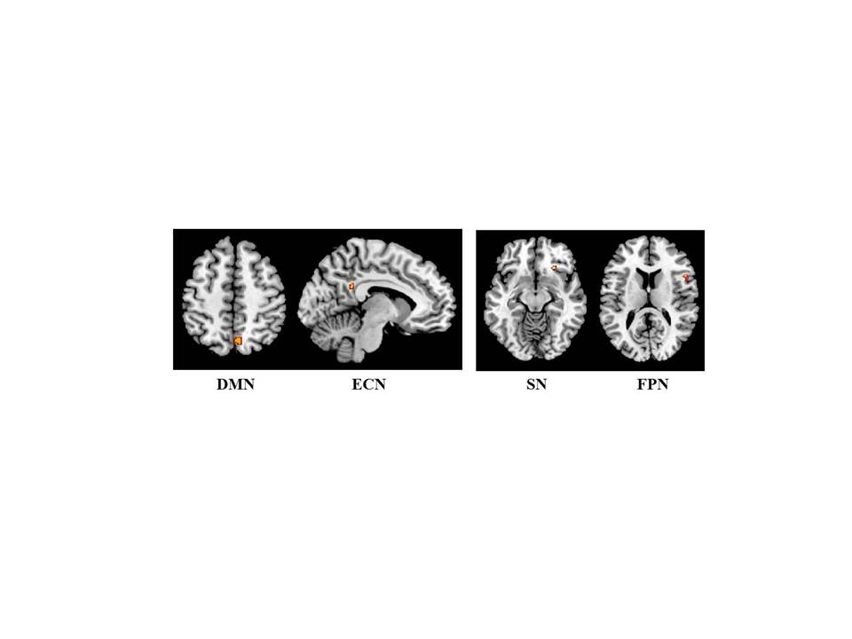
Figure
1. Changes of resting state functional connectivity
in the main cortical functional networks detected in patients with multiple
sclerosis and fatigue after four weeks of treatment with fampridine.
Red-yellow: increased connectivity at follow-up compared to baseline. Abbreviations:
DMV=default mode network; ECN=executive control network; SN=salience network;
FPN=frontoparietal network.
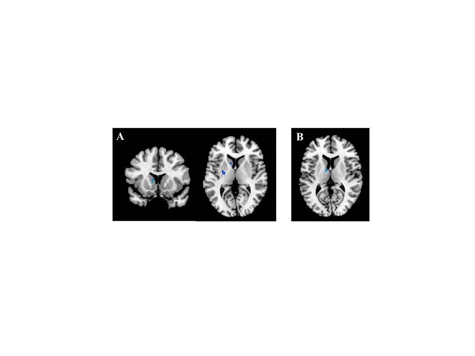
Figure
2. Changes of resting state functional connectivity
in the thalamic network detected in patients with multiple sclerosis and fatigue
after four weeks of treatment with fampridine (A) and amantadine (B). Blue-lightblue:
decreased connectivity at follow-up compared to baseline.