3275
Age related changes in topological properties of brain functional network and structural connectivity1Radiology, Sichuan University, Chengdu, China, 2Sichuan University, Chengdu, China
Synopsis
Title of the abstract was written. The body of the abstract was within word limits. Acknowledgements were mentioned. one figure was uploaded with figure caption.
Abstract
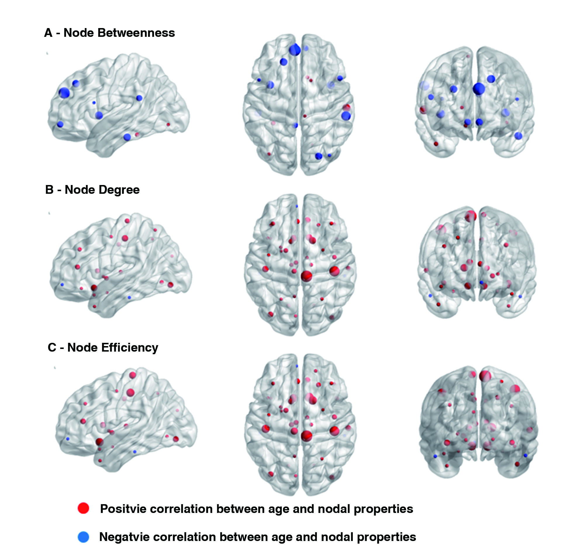
INTRODUCTION: There are still uncertainties about the true nature of age related changes in topological properties of the brain functional network and its structural connectivity during various developmental stages. As structural variability is presumed to be one of the main sources of functional variability expressed in neural dynamics and behavioral performance, evaluation of relationships between structural and functional brain networks in nexus with healthy aging process becomes essential in order to understand the possible mechanisms behind neurodegenerative diseases such as Alzheimer's and Parkinson's. Therefore, in this cross- sectional study, we investigated the effects of age and its relationship with regional nodal properties of the functional brain network and white matter integrity. METHOD: DTI and fMRI data were acquired on a two different (but identical) 3T MRI systems from 458 healthy Chinese participants ranging from age 8 to 81 years. Tractography was conducted on the DTI data using FSL. Graph Theory analyses were conducted on the functional data yielding topological properties of the functional network using SPM and GRETNA toolbox. All statistical analyses were done using SPSS (version 19). Two multiple regressions were performed to investigate the effects of age on nodal topological properties of the functional brain network and white matter integrity. Nodal topological properties and mean FA values were kept as dependent variables, while age, gender and education as independent variables. RESULT: For the functional studies, we observed that regional nodal characteristics such as node betweenness were decreased while node degree and node efficiency was increased in relation to increasing age Perversely, we observed that the relationship between nodal topological properties and fasciculus structures were primarily positive for nodal betweenness but negative for nodal degree and nodal efficiency. Decrease in functional nodal betweenness was primarily located in superior frontal lobe, right occipital lobe and the global hubs On the contrary to the functional studies, we found that the relationship between nodal topological properties and fasciculus structures were primarily positive for nodal betweenness and negative for nodal degree and nodal efficiency. These brain regions also had both direct and indirect anatomical relationships with the 14 fiber bundles. A linear age related decrease in the Fractional anisotropy (FA) value was found in the callosum forceps minor. DISCUSSION: Decrease in nodal betweenness in frontal, right occipital and the global hubs indicates these regions to be most vulnerable with advancing age and might explain the reason behind the cognitive decline in the elderly. A positive relationship between the fasciculus FA and node betweenness in most regions suggests that increased structural connectivity of the fasciculus may improve communication between a node and other nodes in the network. Likewise, both direct and indirect relationships with the 14 fiber bundles might be due to the co-activation of the regions even when there is no direct structural connection between them (1). One of the reasons for not having direct one to one structural and functional connectivity is that they might be mediated through the third party without direct structural connections(2) or it might be due to the false negative results in the tractography analysis or due to false positive resting state fMRI. Our findings of functional connectivity not correlating with structural connectivity is unique in the sense that most of the previous studies have showed that the strength of functional connectivity to be positively correlated with structural connectivity (3, 4, 5, 6). CONCLUSION: These results suggests that age related differences were more pronounced in the functional than in structural measure indicating these measures do not have direct one-to-one mapping. Our study also indicates that the fiber bundles with longer fibers exhibited a more pronounced effect on the properties of functional network.Acknowledgements
This study was supported by the National Natural Science Foundation (Grant Nos. 81222018, 81371527, 81030027, 81227002 and 81220108013), National Key Technologies R&D Program (Program No. 2012BAI01B03) and Program for Changjiang Scholars and Innovative Research Team in University (PCSIRT, Grant No. IRT1272) of China, Dr. Qiyong Gong acknowledges the support from his CMB Distinguished Professorship Award (Award No. F510000/ G16916411), administered by the Institute of International Education, USA.References
1. 1. Honey, C.J., Sporns, O., Cammoun, L., Gigandet, X., Thiran, J.P., Meuli, R., Hagmann, P. (2009) Predicting human resting-state functional connectivity from structural connectivity. Proceedings of the National Academy of Sciences of the United States of America, 106:2035-2040.
2. Greicius, M.D., Supekar, K., Menon, V., Dougherty, R.F. (2009) Resting-state functional connectivity reflects structural connectivity in the default mode network. Cerebral cortex (New York, N.Y. : 1991), 19:72-8.
3. Hagmann, P., Cammoun, L., Gigandet, X., Meuli, R., Honey, C.J., Wedeen, V.J., Sporns, O. (2008) Mapping the structural core of human cerebral cortex. PLoS biology, 6:e159.
4. Honey, C.J., Sporns, O., Cammoun, L., Gigandet, X., Thiran, J.P., Meuli, R., Hagmann, P. (2009) Predicting human resting-state functional connectivity from structural connectivity. Proceedings of the National Academy of Sciences of the United States of America, 106:2035-2040.
5. Koch, M.A., Norris, D.G., Hund-Georgiadis, M. (2002) An investigation of functional and anatomical connectivity using magnetic resonance imaging. NeuroImage, 16:241-50.
6. Skudlarski, P., Jagannathan, K., Calhoun, V.D., Hampson, M., Skudlarska, B.A., Pearlson, G. (2008) Measuring brain connectivity: diffusion tensor imaging validates resting state temporal correlations. NeuroImage, 43:554-61.
Figures