3274
Brain structural plasticity associated with emotional processing in postpartum women: A longitudinal voxel-based morphometry study1Department of Electronic Science, Xiamen University, Xiamen, China, 2Shanghai Key Laboratory of Magnetic Resonance and Department of Physics, School of Physics and Materials Science, East China Normal University, Shanghai, China, 3Department of Special Education, Faculty of Education, East China Normal University, Shanghai, China
Synopsis
Pregnancy constitutes a significant period in women’s lives, after which they often experience numerous physiological and psychological changes. However, structural changes in the brains of postpartum women remain unclear. To investigate these phenomena, we recruited forty-seven postpartum women to participate in a longitudinal magnetic resonance imaging study. Our results first suggest that brain structures in postpartum women show adaptive plasticity, especially regarding alterations in empathy-related regions, including grey matter volume, white matter volume, and cortical thickness, that can facilitate effective adaption and
Introduction
Significant changes in women after delivery may promote the postpartum process of adapting to the new role of motherhood. According to previous studies, the hormones can regulate human social interactions and may further influence the functions and structures of the brain. Fluctuations in hormone levels along with other endocrine changes in women after pregnancy may affect both the function and structure of their brain tissues. Functional neuroimaging studies indicate that new mothers’ brains may develop some adaptive functional changes to the new role of motherhood during pregnancy. Until recently, little was known about brain structural plasticity in relation to pregnancy in humans. In the current longitudinal and noninvasive MRI study, we investigate brain structural plasticity and correlations between brain structural plasticity and empathetic abilities in the postpartum group with a 2-year follow-up using voxel-based morphometry (VBM) analysis.Materials and Methods
We recruited forty-seven postpartum women (age 30.4 ± 2.4) of Chinese ethnicity to participate in this longitudinal voxel-based morphometry study twice two years. Experiments occurred over the course of two scans with a follow-up period (mean time between scan 1 and scan 2: 704 ± 2.0 days). All scans were performed on a Siemens 3.0 T Trio Tim MR system at the Shanghai Key Laboratory of Magnetic Resonance (East China Normal University, Shanghai, China). Anatomical images covering the whole brain were collected using a high-resolution T1-weighted 3-dimensional magnetization-prepared rapid-acquisition gradient-echo pulse sequence. The acquisition parameters were as follows: TR = 2530 ms, TE = 2.34 ms, 256 × 256 matrix, slice = 192 slices, slice thickness = 1.0 mm, and scan duration = 6 min 3 s. In the experimental process, all participants completed the Interpersonal Reactivity Index (IRI) scale that is a questionnaire assessing empathy, respectively. Data were analyzed with voxel-based morphometry using statistical parametric mapping. We further performed region of interest analysis of differential brain regions to investigate relationships between empathy and structural changes in postpartum women.Results
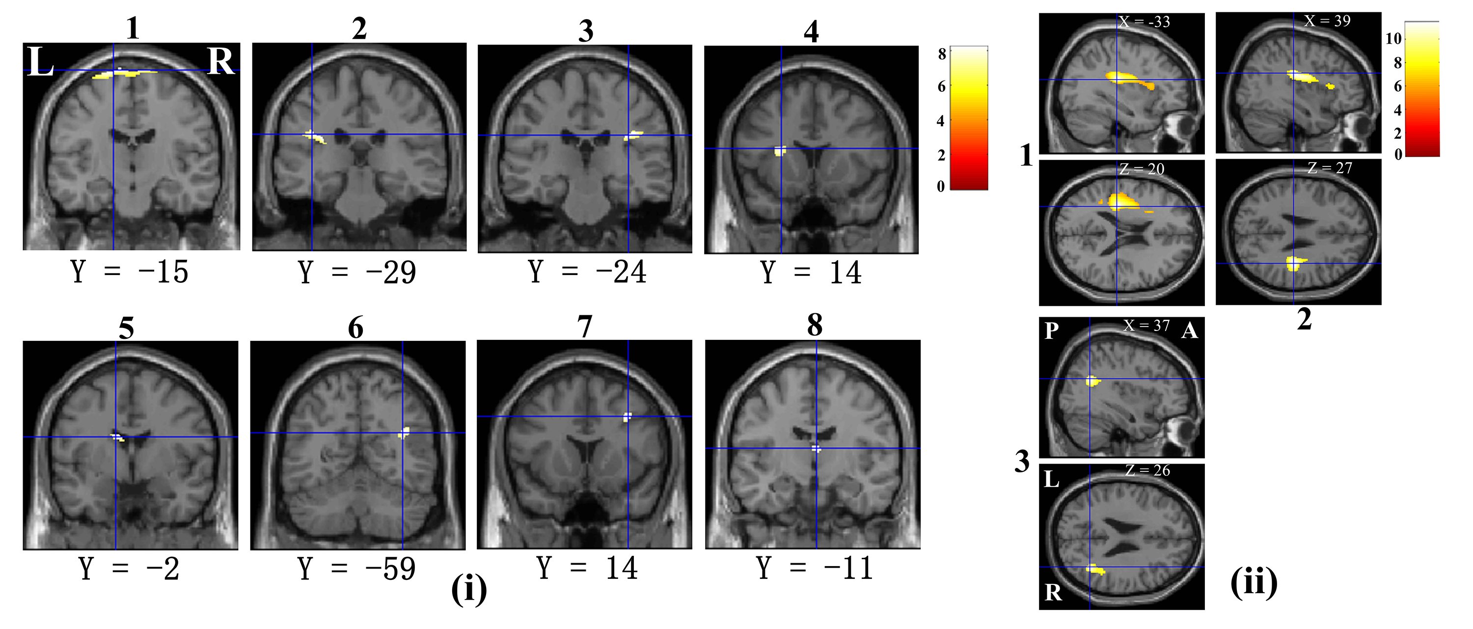
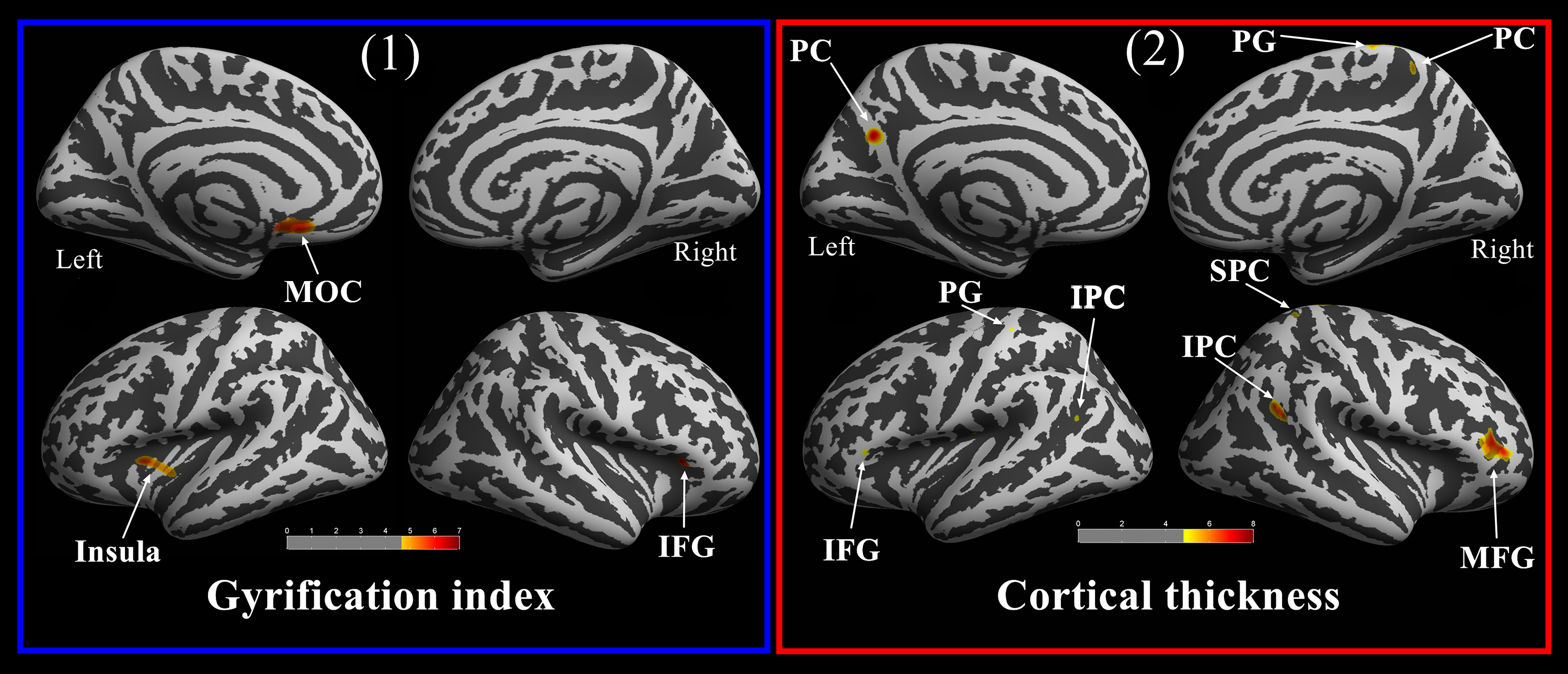
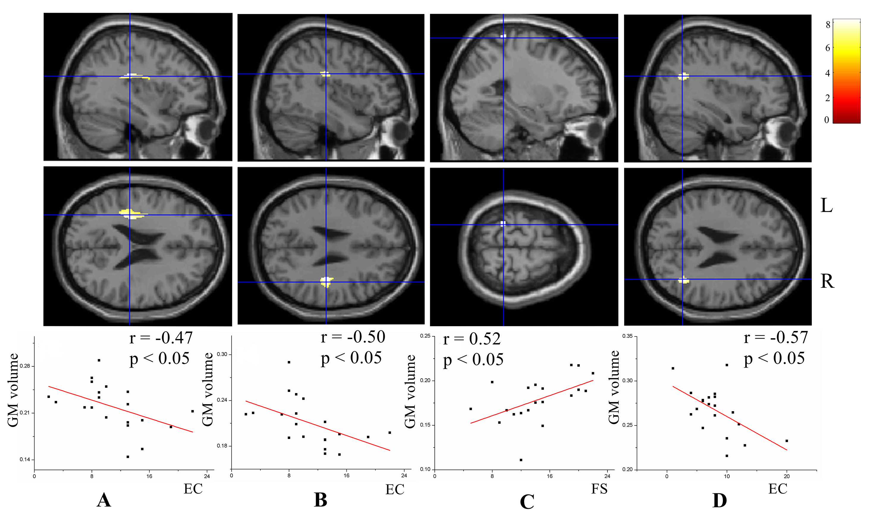
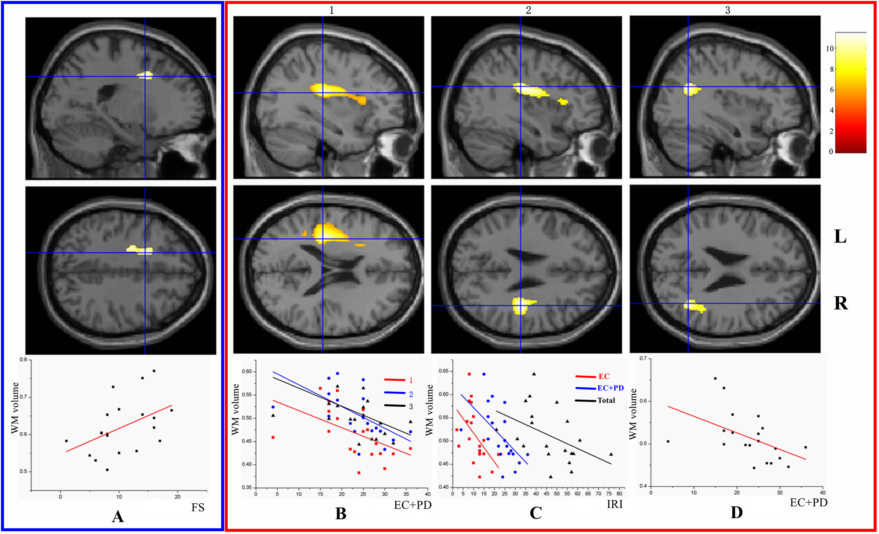
The results reveal that regional grey matter areas associated with cognition and emotional processing are reduced, whereas whiter matter regions exhibit volumetric increases. Surface-based morphometry analysis reflects a reduction in cortical thickness and an increase in cortical gyrification in similar brain regions based on comparisons between the two scans (Figure 1 and Figure 2). Correlation analysis of IRI scores and longitudinal differential grey matter volumes, whiter matter volumes and cortical thickness shows that the empathetic abilities of postpartum women are significantly related to these brain areas (Figure 3 and Figure 4).Discussion
Results are consistent with those of a previous study1 that also found the volume of some GM areas and cortical thickness to be decreased in the brain areas of first-time mothers. Structural plasticity suggests that neurons in the brains of postpartum women are further pruned for specialization and adaptability to better nurture infants2. The observed changes in postpartum women are all functionally correlated with emotional processing and cognitive function. Correlation analysis suggests that a specific relationship between alterations in empathy-related regions, including changes in GM volume, WM volume, and cortical thickness, and empathetic abilities could reflect cognitive and emotional function reorganization associated with parental behaviour neurodevelopment during pregnancy and the postpartum phase. Consequently, results predicted that the brains of postpartum women exhibit structural dynamic plasticity for the adaptive changes in cognitive function and empathic processing. These findings provide a neuroanatomical basis for understanding the processing of emotional sensory information in postpartum women.Acknowledgements
This work was supported by the National Natural Science Foundation of China (grants 81571658 and U1632274) and the Social Science Foundation of China (grant 15ZDB016).References
1. Hoekzema E, et al. Pregnancy leads to long-lasting changes in human brain structure. Nat Neurosci 2017; 20(2): 287-96.
2. Herting MM, et al. A longitudinal study: Changes in cortical thickness and surface area during pubertal maturation. Plos One 2015; 10(3).
Figures