3273
Multimodal study of hyperbaric oxygenation effects on normal human brain metabolism and functional connectivity at 3 Tesla.10501, Emanuel Institute of Biochemical Physics of the Russian Academy of Sciences, Moscow, Russian Federation, 2Radiology, Clinical and Research Institute of Emergent Pediatric Surgery and Trauma, Moscow, Russian Federation, 3National Research Nuclear University MEPhI, Moscow, Russian Federation, 4Dmitry Rogachev National Research Center of Pediatric Hematology, Oncology and Immunology, Moscow, Russian Federation, 5Hyperbaric Oxygen Dept., Clinical and Research Institute of Emergent Pediatric Surgery and Trauma, Moscow, Russian Federation, 6Semenov Institute of Chemical Physics of the Russian Academy of Sciences, Moscow, Russian Federation
Synopsis
This study is aimed to reveal the effects of hyperbaric oxygenation (HBO) on human brain metabolites using 1H and 31P MRS, and functional connectivity using resting-state fMRI. The MRS and rs-fMRI studies were performed twice: before and after one HBO session (p=1.2 atmosphere, 100% O2). The results demonstrate the positive effect of hyperbaric oxygenation on human brain metabolism and functioning.
Introduction
One of perspective ways of hypoxia-caused disorders treatment is hyperbaric oxygenation (HBO). The clinical and laboratory studies have demonstrated its effectiveness in different cases, for example, in CO poisoning [1]. The in vivo studies of HBO influence on metabolism might be of interest; previously we have demonstrated increase in α-ATP peak intensity [2]. The aim of this study is to reveal the effect of one low-pressure (1.2 atmosphere absolute, ATA) HBO session on human brain metabolism using 1H and 31P MRS, and functional connectivity using resting-state fMRI.Materials and methods
Philips Achieva 3.0T, Philips Head SENSE coil, 31P/1H bird-cage coil (Rapid Biomed), hyperbaric chamber Sechrist 3200 were used.
Seventeen normal subjects (18-30 years old) participated in 31P MRS part of study and 6 normals (20-24 years old) – in 1H MRS and rs-fMRI part of study. Both parts of the study followed the plan: 1) MRI+MRS; 2) HBO session; 3) repeat of MRI+MRS.
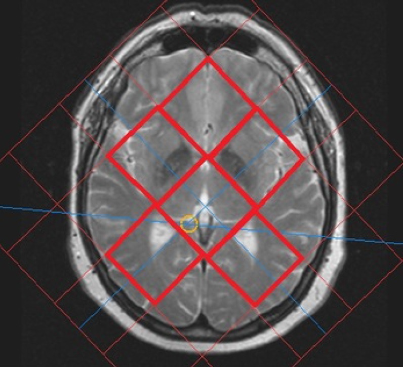
31P MRS part: spectra were obtained after axial and sagittal (2 minutes) T2-weighted images. Voxels location is demonstrated on figure 1. The parameters of 2D 31P MRS are as follows: pulse sequence ISIS, TE = 0.3 ms, TR = 1900 ms, NSA = 16, FA = 35°, broadband NOE and decoupling, slice thickness = 30 mm, FOV = 200x200 mm, voxel size = 40x40 mm.
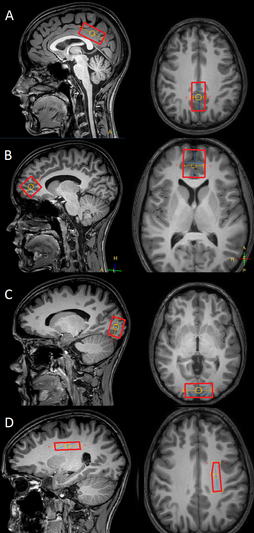
1H MRS and rs-fMRI part: resting state fMRI (GE-EPI, TR=3000 ms, TE=30 ms, EPI factor = 39, slice thickness 3 mm, matrix size = 112x116, 80 dynamic scans) and 4 single-voxel 1H MRS (PRESS, TE = 115 ms, TR = 2000 ms) were obtained after 3D T1 images. 1H MRS voxels are located in medial prefrontal cortex (25x25x25 mm), posterior cingular cortex (40x20x20 mm), visual cortex (20x40x30 mm) and left subcortical white matter (35x10x10 mm), the location is demonstrated on fig.2.
The HBO session lasted for 50 minutes (5 minutes for achieving constant 1.2 atmospheres mode, 40 minutes at the mode and 5 minutes for decompression). The time required for a patient to get into MRI scanner after HBO did not exceed 5 minutes.
Data processing
The 2D 31P MRS data processing was performed in jMRUI 5.2. The FIDs of voxels that were fully located in brain (red frames on fig.1) were averaged, two spectra remained for each subject: before and after HBO session. Integral intensities of spectral peaks were quantified using AMARES algorithm. For each subject in spectra before and after HBO session the intensities of individual peaks were normalized to the sum of integral intensities of all peaks. pH value before and after HBO session was calculated from PCr-Pi chemical shift difference. Relative values (after HBO/before HBO) of all parameters were calculated.
Single voxel 1H MRS data was processed in LCModel. Metabolite concentrations (relative to unsuppressed H2O) in spectra from different locations after HBO session were normalized to the according concentrations in spectra before HBO.
Statistical processing was performed in STATISTICA 12.
The rs-fMRI data was processed in CONN (Matlab). The correlation coefficients of Default Mode Network nodes with different anatomical structures after HBO session were in case of statistical significance (p-FDR<0.05) normalized on the according coefficients before HBO.
Results
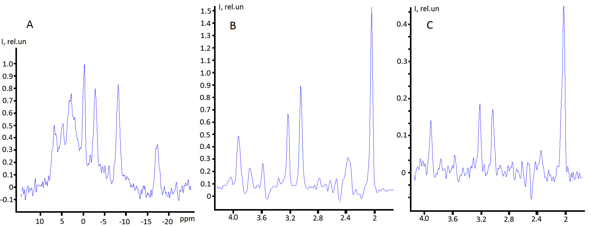
Typical spectra obtained in this study are demonstrated on figure 3.
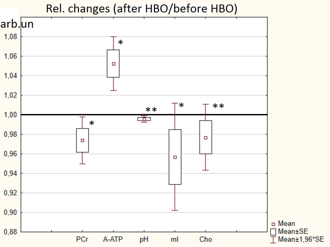
The effects of HBO session in whole brain according to 31P and 1H MRS data: α-ATP increased (~5%, p<0.01), PCr decreased (~3%, p<0.01), pH decreased (~0.5%, p<0.05), myo-inositol decreased (~4%, p<0.01), Cho decreased (~2%, p<0.05). Relative changes in comparison with value=1 are demonstrated on figure 4. Other 31P and 1H MRS parameters remained unchanged.
The correlation coefficients (beta) of Medial PreFrontal Cortex with Frontal Medial Cortex and Cingulate Gyrus increased (p-FDR<0.05) by ~10% after HBO session according to rs-fMRI data.
Discussion
The results obtained demonstrate direct activation of energy metabolism by a HBO session. As we discussed before [2], since γ- and β-ATP remain unchanged and uridine-diphosphate glucose at -9.72 ppm [3] cannot be picked out of noise neither in spectrum before nor after HBO session, the increase of α-ATP peak integral intensity means [NAD] increase. Updated results in current work indirectly confirm this fact, since PCr and pH decrease may witness for energy-consuming synthesis of NAD from Nicotinamide Mononucleotide [4].
The decrease of choline phenomenon as well as decrease of myo-inositol hence to one HBO session remains a subject of future research.
Nonetheless, we have demonstrated the metabolism activation, NAD synthesis and increased correlation of functional connectivity in the zones responsible for cognitive functions. The results confirm the HBO effectiveness for human brain stimulation even at low pressures (p=1.2 atmospheres).
Acknowledgements
No acknowledgement found.References
1. G.G. Rogatsky et al. Hyperbaric oxygenation affects rat brain function after carbon monoxide exposure. UHM 2002, Vol 29, No. 1
2. Manzhurtsev et. al. Electronic poster at ISMRM 2017, http://dev.ismrm.org/2017/5610.html
3. Ren et. al. NMR Biomed. 2015; 28: 1455–1462 DOI: 10.1002/nbm.3384
4. Long et al. BMC Neurology (2015) 15:19 DOI 10.1186/s12883-015-0272-x
Figures