3272
Altered Topological Properties of Brain Functional Networks in Type I GD Patients During a Near Five-year Follow-upDi Hu1, Miao Zhang1, Shengpei Wang2, Huiying Kang1, and Yun Peng1
1Beijing Children's Hospital, Capital Medical University,National Center for Children's Health, China, Beijing, China, 2State Key Laboratory of Management and Control for ComplexSystems, Institute of Automation, Chinese Academy of Sciences, Beijing, China, Beijing, China
Synopsis
Our previous had shown altered nodal properties within the executive control network in children with type I GD. This time we further investigate the variation trend of brain topological properties in type I GD patients during a near five-year folloe-up under regular enzyme replacement. Results showed that the efficiency of functional segregation in brain network were increased and more nodes were involved and reorganized. The result may explain the improved mood and global functioning that have previous reported.
Introduction
Our previous study had shown altered nodal properties within the executive control networkin children with type I GD. And some studies confirmed that mood and global functioning showed significant improvement and psychological symptoms were fewer than before after a period of regular enzyme replacement therapy. But fewer studies have investigated the variation trend of brain topological properties in type I GD patients under regular enzyme replacement therapy(ERT) . So our study aims to explore this question.Methods
Resting-state functional MRI scans were obtained from sixteen type I GD patients before and after a near five-year period with regular ERT. Utilizing graph-based network analysis, we compared alteration of topological properties of brain functional networks at global and nodal levels within them. Paried t test with false discovery rate correction was performed to investigate the differences. Pearson correlation analyses were carried out to test the relationships between clinical parameters and altered network properties.Results
During a near five-year follow-up, the type I GD patients showed significantly increased local efficiency and normalized clustering coefficient () of the global network properties in some specific thresholds(p < 0.05, FDR corrected). Nine ROIs involving eight regions which included right inferior frontal gyrus, right superior parietal gyrus, pecuneus and left middle/inferior frontal Gyrus, inferior parietal sulcus, precuneus, thalamus showed increased nodal efficiency. Meanwhile five ROIs involving five regions which included right inferior frontal gyrus, superior parietal gyrus, precuneus and left middle frontal gyrus, thalamus showed increased nodal degree. On the contrary, two ROIs involving three regions which included right crus I, VIII and Vllb showed decreased nodal degree. And one ROI which included crus VIII and Vllb showed decreased nodal efficiency. The altered ROIs were important component of the executive control network, salience network, default mode network, basal ganglia network and visuospatial network. No significant alterations of betweenness centrality values were found in all the hubs. Unfortunately, no correlations were found between the altered nodal efficiency and the clinical parameters.Conclusion
We are the first to investigate the alteration trend of the functional brain network in type I GD patients. After a near five-year follow-up, these patient showed increased efficiency of functional segregation in brain network and more diffuse involved and reorganized nodes, which may to some extent explained the improvement in mood and global functioning and the less psychological symptoms. And we believe that our results provide new insight to further understand the neurophysiological change of the type I GD patients.Acknowledgements
No acknowledgement found.References
No reference found.Figures
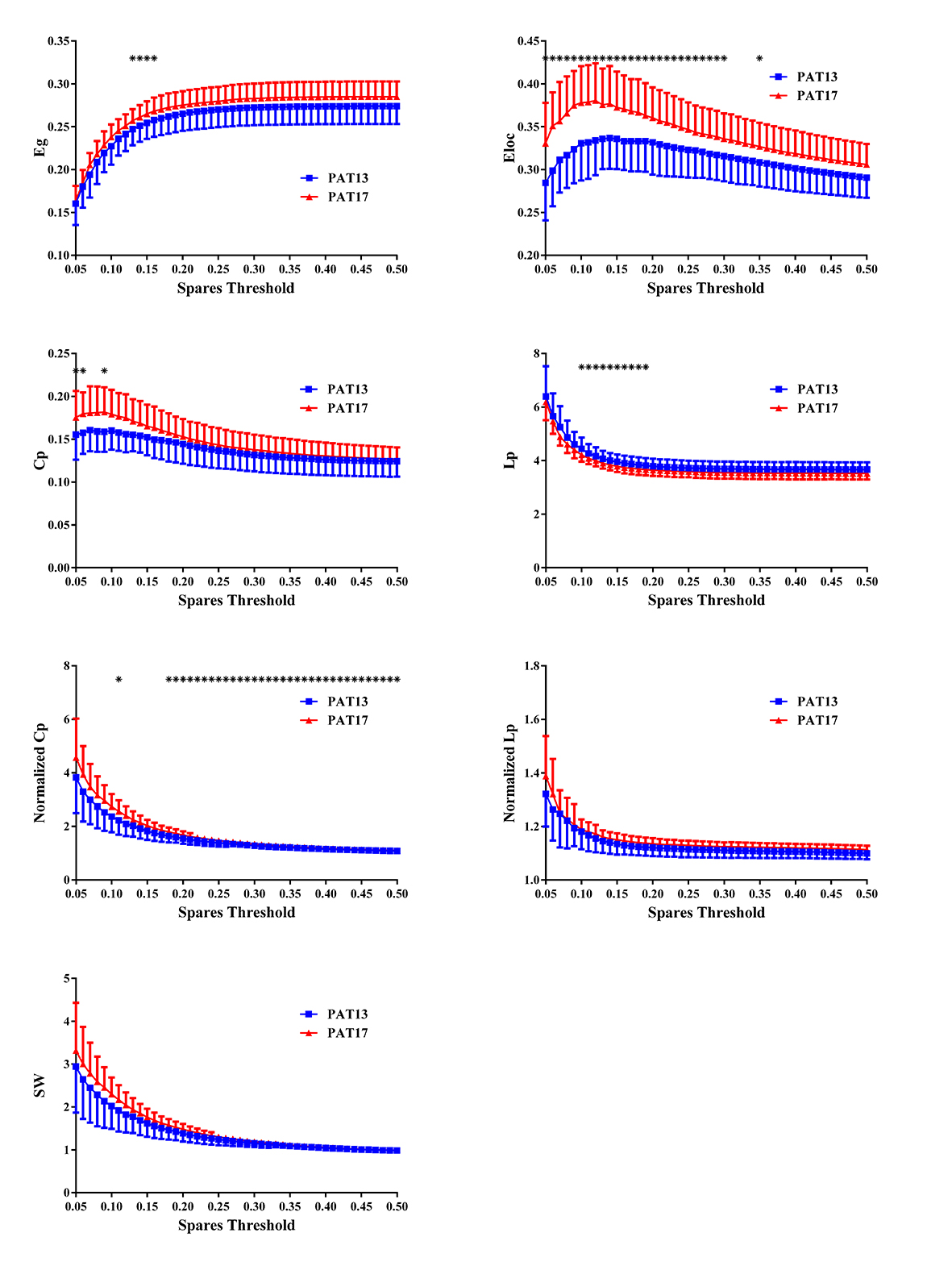
Figure 1. Global graph theoretical measurements in type I GD patients between before and after a near five-year follow-up. * means significant between-group differences. CON: normal controls. PAT: type I GD patients
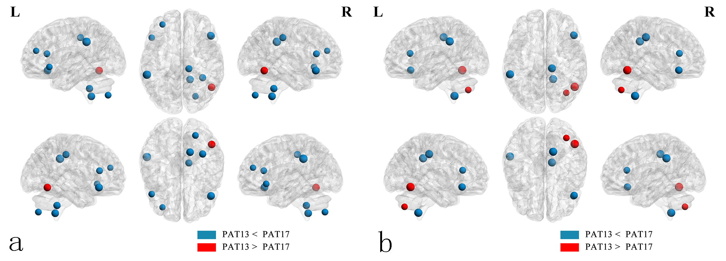
Figure 2. (a) and (b) show altered nodes representing mean nodal efficiency and degree separately in the type I GD patients before and after the five-year follow-up. Green represents decreased nodal properties. Red represents increased nodal properties.