3271
Disrupted Nodal Organization of brain functional Network in Children with Type I Gaucher Disease1Beijing Children's Hospital, Capital Medical University,National Center for Children's Health, China, Beijing, China, 2State Key Laboratory of Management and Control for ComplexSystems, Institute of Automation, Chinese Academy of Sciences, Beijing, China, Beijing, China
Synopsis
In our study, we utilize graph-based network analysis to investigate the topological properties of resting-state brain functional connectivity in children with type I GD. Sixteen children diagnosed as type I GD and sixteen age- and sex- matched healthy controls were recruited. No alterations were found at global level. Three nodes within the executive control network showed decreased nodal degree or efficiency. Our provide new insight to further understand the neurophysiological change of these patients.
Introduction
As an autosomal recessive glycosphingolipid storage disease, many studies have investigated the brain structural abnormalities in children with type I GD. But little is known about their brain functional network. So our aim is to investigate the topological properties of resting-state brain functional connectivity in these patients.Methods
Sixteen children diagnosed as type I GD ranging from 7.7to17.6 years old and sixteen age- and sex- matched healthy controls were recruited in our study. All of them underwent rs-fMRI examination. Utilizing graph-based network analysis, we compared topological properties of brain functional networks between children with type I GD and the healthy controls. Two-sample t test with false discovery rate correction was performed to investigate the differences. Pearson correlation analyses were carried out to test the relationships between clinical parameters and altered network properties.Results
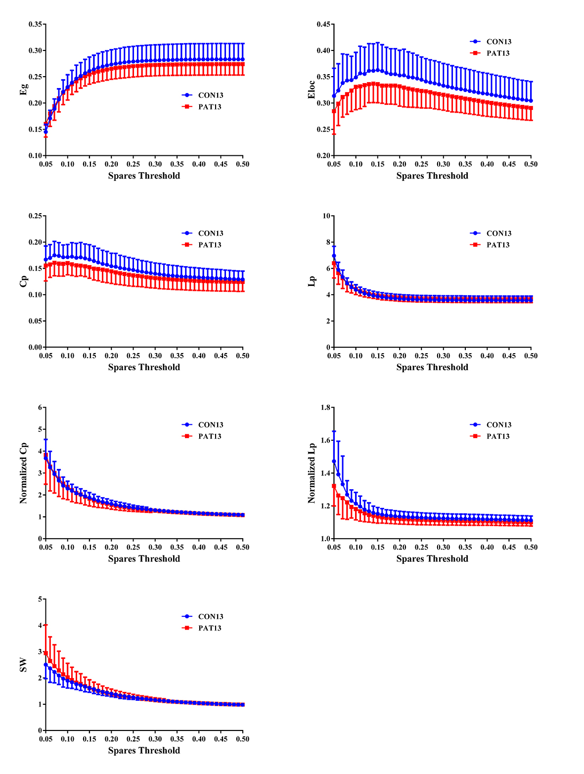
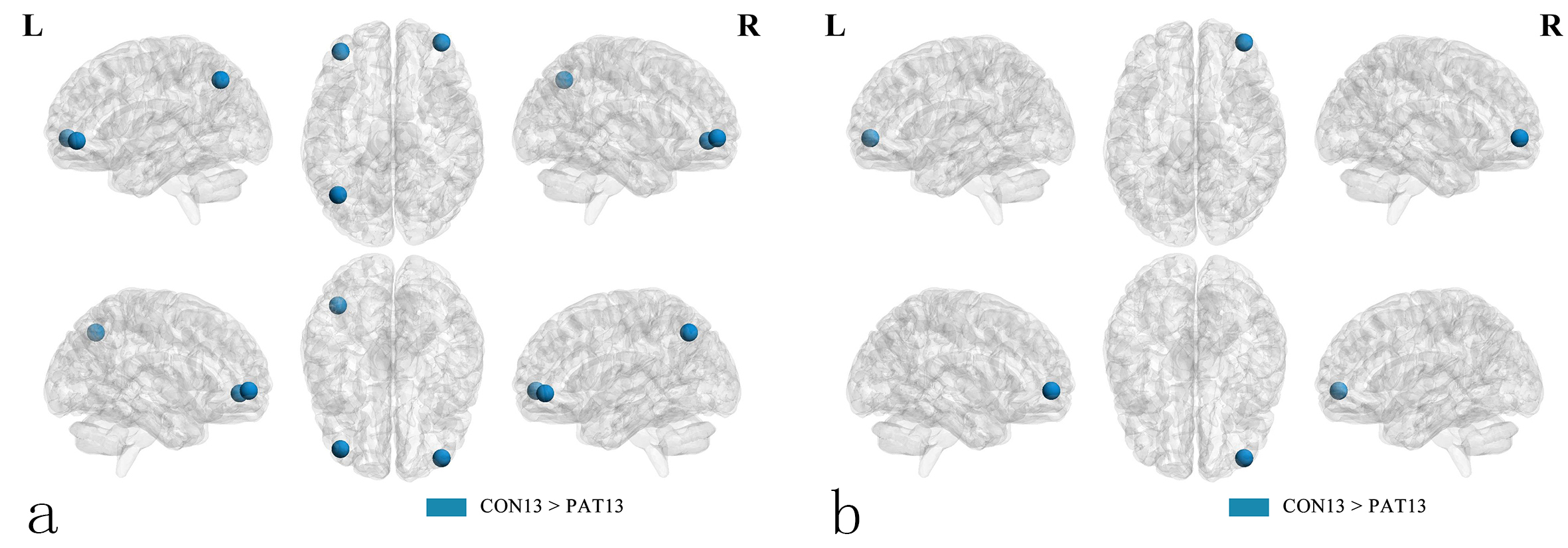
Compared with the healthy controls, the type I GD patients showed no significant difference in global network properties(mean degree, global efficiency, local efficiency, Cp, Lp, , and σ over the wide range of thresholds(Figure 1). Three nodes involving seven brain regions within the executive control network showed decreased mean nodal efficiency. Meanwhile the right middle frontal gyrus among them also showed decreased nodal degree(Figure 2). No significant alterations of betweenness centrality values were found in all the hubs. Unfortunately, no correlations were found between the altered nodal efficiency and the clinical parameters.Conclusion
We are the first to investigate the functional brain network in children with type I GD. And our results provide new insight to further understand the neurophysiological change of these patients, which may result from the brain infiltration of GD cells.Acknowledgements
No acknowledgement found.References
[1] Abdel R A, Abd E N, Abdalla A, et al. Apparent diffusion coefficient vale of the brain in patients with Gaucher's disease type II and type III[J]. Neuroradiology,2009,51(11):773-779.
[2] Davies E H, Seunarine K K, Banks T, et al. Brain white matter abnormalities in paediatric Gaucher Type I and Type III using diffusion tensor imaging[J]. J Inherit Metab Dis,2011,34(2):549-553.
Figures